Low PA, Opfer-Gehrking TL, McPhee BR, Fealey RD, Benarroch EE, Willner CL, et al. Prospective evaluation of clinical characteristics of orthostatic hypotension. Mayo Clin Proc. 1995. 70:617-622. [QxMD MEDLINE Link].
LASSEN NA. Cerebral blood flow and oxygen consumption in man. Physiol Rev. 1959. 39:183-238. [QxMD MEDLINE Link].
Ocon AJ, Medow MS, Taneja I, Clarke D and Stewart JM. Decreased upright cerebral blood flow and cerebral autoregulation in normocapnic postural tachycardia syndrome. Am J Physiol Heart Circ Physiol. 2009. 297:H664-H673. [QxMD MEDLINE Link].
Ocon AJ, Kulesa J, Clarke D, Taneja I, Medow MS and Stewart JM. Increased phase synchronization and decreased cerebral autoregulation during fainting in the young. Am J Physiol Heart Circ Physiol. 2009. 297:H2084-H2095. [QxMD MEDLINE Link].
Del Pozzi AT, Schwartz CE, Tewari D, Medow MS and Stewart JM. Reduced cerebral blood flow with orthostasis precedes hypocapnic hyperpnea, sympathetic activation, and postural tachycardia syndrome. Hypertension. 2014. 63:1302-1308. [QxMD MEDLINE Link].
Lagi A, Cencetti S, Corsoni V, Georgiadis D and Bacalli S. Cerebral vasoconstriction in vasovagal syncope: any link with symptoms? A transcranial Doppler study. Circulation. 2001. 104:2694-2698. [QxMD MEDLINE Link].
Stewart JM, Medow MS, Cherniack NS and Natelson BH. Postural hypocapnic hyperventilation is associated with enhanced peripheral vasoconstriction in postural tachycardia syndrome with normal supine blood flow. Am J Physiol Heart Circ Physiol. 2006. 291:H904-H913. [QxMD MEDLINE Link].
Hamel E. Perivascular nerves and the regulation of cerebrovascular tone. J Appl Physiol (1985 ). 2006. 100:1059-1064. [QxMD MEDLINE Link].
Freeman R, Wieling W, Axelrod FB, Benditt DG, Benarroch E, Biaggioni I, et al. Consensus statement on the definition of orthostatic hypotension, neurally mediated syncope and the postural tachycardia syndrome. Clin Auton Res. 2011. 21:69-72. [QxMD MEDLINE Link].
Shibao C, Okamoto L and Biaggioni I. Pharmacotherapy of autonomic failure. Pharmacol Ther. 2012. 134:279-286. [QxMD MEDLINE Link].
Hilz MJ, Ehmann EC, Pauli E, Baltadzhieva R, Koehn J, Moeller S, et al. Combined counter-maneuvers accelerate recovery from orthostatic hypotension in familial dysautonomia. 2012. 126:162-170. [QxMD MEDLINE Link].
Robertson D, Haile V, Perry SE, Robertson RM, Phillips JA, III and Biaggioni I. Dopamine beta-hydroxylase deficiency. A genetic disorder of cardiovascular regulation. Hypertension. 1991. 18:1-8. [QxMD MEDLINE Link].
Klein CM, Vernino S, Lennon VA, Sandroni P, Fealey RD, Benrud-Larson L, et al. The spectrum of autoimmune autonomic neuropathies. Ann Neurol. 2003. 53:752-758. [QxMD MEDLINE Link].
Singh NK, Jaiswal AK, Misra S and Srivastava PK. Assessment of autonomic dysfunction in Guillain-Barre syndrome and its prognostic implications. Acta Neurol Scand. 1987. 75:101-105. [QxMD MEDLINE Link].
Mukai S and Lipsitz LA. Orthostatic hypotension. Clin Geriatr Med. 2002. 18:253-268. [QxMD MEDLINE Link].
Jain S and Goldstein DS. Cardiovascular dysautonomia in Parkinson disease: from pathophysiology to pathogenesis. Neurobiol Dis. 20122012. 46:572-580. [QxMD MEDLINE Link].
Furlan R, Jacob G, Snell M, Robertson D, Porta A, Harris P and Mosqueda-Garcia R. Chronic orthostatic intolerance: a disorder with discordant cardiac and vascular sympathetic control. Circulation. 1998. 98:2154-2159. [QxMD MEDLINE Link].
Schondorf R and Low PA. Idiopathic postural orthostatic tachycardia syndrome: an attenuated form of acute pandysautonomia?. Neurology. 1993. 43:132-137. [QxMD MEDLINE Link].
Singer W, Sletten DM, Opfer-Gehrking TL, Brands CK, Fischer PR and Low PA. Postural tachycardia in children and adolescents: what is abnormal?. J Pediatr. 2012. 160:222-226. [QxMD MEDLINE Link].
Stewart JM, Glover JL and Medow MS. Increased plasma angiotensin II in postural tachycardia syndrome (POTS) is related to reduced blood flow and blood volume. Clin Sci (Lond). 2006. 110:255-263. [QxMD MEDLINE Link].
Jacob G, Costa F, Shannon JR, Robertson RM, Wathen M, Stein M, et al. The neuropathic postural tachycardia syndrome. N Engl J Med. 2000. 343:1008-1014. [QxMD MEDLINE Link].
Stewart JM. Pooling in chronic orthostatic intolerance: arterial vasoconstrictive but not venous compliance defects. Circulation. 2002. 105:2274-2281. [QxMD MEDLINE Link].
Stewart JM, Medow MS, Glover JL and Montgomery LD. Persistent splanchnic hyperemia during upright tilt in postural tachycardia syndrome. Am J Physiol Heart Circ Physiol. 2006. 290:H665-H673. [QxMD MEDLINE Link].
Stewart JM, Munoz J and Weldon A. Clinical and physiological effects of an acute alpha-1 adrenergic agonist and a beta-1 adrenergic antagonist in chronic orthostatic intolerance. Circulation. 2002. 106:2946-2954. [QxMD MEDLINE Link].
Stewart JM, Rivera E, Clarke DA, Baugham IL, Ocon AJ, Taneja I, et al. Ventilatory baroreflex sensitivity in humans is not modulated by chemoreflex activation. Am J Physiol Heart Circ Physiol. 2011. 300:H1492-H1500. [QxMD MEDLINE Link].
Bonyhay I and Freeman R. Sympathetic nerve activity in response to hypotensive stress in the postural tachycardia syndrome. Circulation. 2004. 110:3193-3198. [QxMD MEDLINE Link].
Shannon JR, Flattem NL, Jordan J, Jacob G, Black BK, Biaggioni I, et al. Orthostatic intolerance and tachycardia associated with norepinephrine-transporter deficiency. N Engl J Med. 2000. 342:541-549. [QxMD MEDLINE Link].
Lambert E, Eikelis N, Esler M, Dawood T, Schlaich M, Bayles R, et al. Altered sympathetic nervous reactivity and norepinephrine transporter expression in patients with postural tachycardia syndrome. Circ Arrhythm Electrophysiol. 2008. 1:103-109. [QxMD MEDLINE Link].
Zanzinger J, Czachurski J and Seller H. Neuronal nitric oxide reduces sympathetic excitability by modulation of central glutamate effects in pigs. Circ Res. 1997. 80:565-571. [QxMD MEDLINE Link].
Storgaard T and Nedergaard OA. Prejunctional modulation by angiotensins of noradrenaline release from sympathetic neurons in isolated rabbit aorta. Naunyn Schmiedebergs Arch Pharmacol. 1997. 356:706-711. [QxMD MEDLINE Link].
Kolo LL, Westfall TC and Macarthur H. Nitric oxide decreases the biological activity of norepinephrine resulting in altered vascular tone in the rat mesenteric arterial bed. Am J Physiol Heart Circ Physiol. 2004. 286:H296-H303. [QxMD MEDLINE Link].
Hatanaka Y, Zamami Y, Koyama T, Hobara N, Jin X, Kitamura Y and Kawasaki H. A ketolide antibiotic, telithromycin, inhibits vascular adrenergic neurotransmission in the rat mesenteric vascular bed. Br J Pharmacol. 2008. 155:826-836. [QxMD MEDLINE Link].
Macarthur H, Mattammal MB and Westfall TC. A new perspective on the inhibitory role of nitric oxide in sympathetic neurotransmission. Biochem Biophys Res Commun. 1995. 216:686-692. [QxMD MEDLINE Link].
Hu ZW, Shi XY, Okazaki M and Hoffman BB. Angiotensin II induces transcription and expression of alpha 1-adrenergic receptors in vascular smooth muscle cells. Am J Physiol. 1995. 268:H1006-H1014. [QxMD MEDLINE Link].
Head GA. Role of AT1 receptors in the central control of sympathetic vasomotor function. Clin Exp Pharmacol Physiol. 1996. Suppl 3:S93-S98. [QxMD MEDLINE Link].
Campese VM, Ye S, Zhong H, Yanamadala V, Ye Z and Chiu J. Reactive oxygen species stimulate central and peripheral sympathetic nervous system activity. Reactive oxygen species stimulate central and peripheral sympathetic nervous system activity. 2004. 287:H695-H703. [QxMD MEDLINE Link].
Wolin MS. Interactions of oxidants with vascular signaling systems. Arterioscler Thromb Vasc Biol. 2000. 20:1430-1442. [QxMD MEDLINE Link].
Landmesser U, Dikalov S, Price SR, McCann L, Fukai T, Holland SM, et al. Oxidation of tetrahydrobiopterin leads to uncoupling of endothelial cell nitric oxide synthase in hypertension. J Clin Invest. 2003. 111:1201-1209. [QxMD MEDLINE Link].
Raj SR, Biaggioni I, Yamhure PC, Black BK, Paranjape SY, Byrne DW and Robertson D. Renin-aldosterone paradox and perturbed blood volume regulation underlying postural tachycardia syndrome. Circulation. 2005. 111:1574-1582. [QxMD MEDLINE Link].
Medow MS, Bamji N, Clarke D, Ocon AJ and Stewart JM. Reactive oxygen species (ROS) from NADPH and xanthine oxidase modulate the cutaneous local heating response in healthy humans. J Appl Physiol (1985 ). 2011. 111:20-26. [QxMD MEDLINE Link].
Stewart JM, Ocon AJ, Clarke D, Taneja I and Medow MS. Defects in cutaneous angiotensin-converting enzyme 2 and angiotensin-(1-7) production in postural tachycardia syndrome. Hypertension. 2009. 53:767-774. [QxMD MEDLINE Link].
Gowers WR. A Lecture on Vagal and Vasovagal Attacks. Lancet. 1907. 1551-1554.
Lewis T. A Lecture on VASOVAGAL SYNCOPE AND THE CAROTID SINUS MECHANISM. Br Med J. 1932. 1:873-876. [QxMD MEDLINE Link].
Vaddadi G, Guo L, Esler M, Socratous F, Schlaich M, Chopra R, et al. Recurrent postural vasovagal syncope: sympathetic nervous system phenotypes. Circ Arrhythm Electrophysiol. 2011. 4:711-718. [QxMD MEDLINE Link].
Soteriades ES, Evans JC, Larson MG, Chen MH, Chen L, Benjamin EJ and Levy D. Incidence and prognosis of syncope. N Engl J Med. 2002. 347:878-885. [QxMD MEDLINE Link].
Brignole M and Menozzi C. The natural history of carotid sinus syncope and the effect of cardiac pacing. Europace. 2011. 13:462-464. [QxMD MEDLINE Link].
Engelhardt W, Kotlarek F and von BG. Deglutition syncope in childhood with complete atrioventricular block. Am J Cardiol. 1986. 58:1113-1114. [QxMD MEDLINE Link].
Kapoor WN, Peterson J and Karpf M. Defecation syncope. A symptom with multiple etiologies. Arch Intern Med. 1986. 146:2377-2379. [QxMD MEDLINE Link].
Bae MH, Kang JK, Kim NY, Choi WS, Kim KH, Park SH, et al. Clinical characteristics of defecation and micturition syncope compared with common vasovagal syncope. Pacing Clin Electrophysiol. 2012. 35:341-347. [QxMD MEDLINE Link].
Benditt DG, Samniah N, Pham S, Sakaguchi S, Lu F, Lurie KG and Ermis C. Effect of cough on heart rate and blood pressure in patients with "cough syncope". Heart Rhythm. 2005. 2:807-813. [QxMD MEDLINE Link].
Evans WN, Acherman R, Kip K and Restrepo H. Hair-grooming syncope in children. Clin Pediatr (Phila). 2009. 48:834-836. [QxMD MEDLINE Link].
Sarrigiannis PG, Randall M, Kandler RH, Grunewald RA, Harkness K and Reuber M. Stretch syncope: reflex vasodepressor faints easily mistaken for epilepsy. Epilepsy Behav. 2011. 20:450-453. [QxMD MEDLINE Link].
Colivicchi F, Ammirati F, Biffi A, Verdile L, Pelliccia A and Santini M. Exercise-related syncope in young competitive athletes without evidence of structural heart disease. Clinical presentation and long-term outcome. Eur Heart J. 2002. 23:1125-1130. [QxMD MEDLINE Link].
Moya A, Sutton R, Ammirati F, Blanc JJ, Brignole M, Dahm JB, et al. Guidelines for the diagnosis and management of syncope (version 2009). Eur Heart J. 2009. 30:2631-2671. [QxMD MEDLINE Link].
Aviado DM and Guevara AD. The Bezold-Jarisch reflex. A historical perspective of cardiopulmonary reflexes. Ann N Y Acad Sci. 2001. 940:48-58. [QxMD MEDLINE Link].
Hainsworth R. Syncope: what is the trigger?. Heart. 2003. 89:123-124. [QxMD MEDLINE Link].
Oberg B and Thoren P. Increased activity in left ventricular receptors during hemorrhage or occlusion of caval veins in the cat. A possible cause of the vaso-vagal reaction. Acta Physiol Scand. 1972. 85:164-173. [QxMD MEDLINE Link].
Scherrer U, Vissing S, Morgan BJ, Hanson P and Victor RG. Vasovagal syncope after infusion of a vasodilator in a heart-transplant recipient. N Engl J Med. 1990. 322:602-604. [QxMD MEDLINE Link].
Liu JE, Hahn RT, Stein KM, Markowitz SM, Okin PM, Devereux RB and Lerman BB. Left ventricular geometry and function preceding neurally mediated syncope. Circulation. 2000. 101:777-783. [QxMD MEDLINE Link].
Mosqueda-Garcia R, Furlan R, Tank J and Fernandez-Violante R. The elusive pathophysiology of neurally mediated syncope. Circulation. 2000. 102:2898-2906. [QxMD MEDLINE Link].
Barcroft H and McMichael JES-SEP. Posthaemorrhagic fainting. Study by cardiac output and forearm flow. Lancet. 1944. 243:489-491.
Julien C. The enigma of Mayer waves: Facts and models. Cardiovasc Res. 2006. 70:12-21. [QxMD MEDLINE Link].
Hammer PE and Saul JP. Resonance in a mathematical model of baroreflex control: arterial blood pressure waves accompanying postural stress. Am J Physiol Regul Integr Comp Physiol. 2005. 288:R1637-R1648. [QxMD MEDLINE Link].
Cooke WH, Rickards CA, Ryan KL, Kuusela TA and Convertino VA. Muscle sympathetic nerve activity during intense lower body negative pressure to presyncope in humans. J Physiol. 2009. 587:4987-4999. [QxMD MEDLINE Link].
Taneja I, Medow MS, Glover JL, Raghunath NK and Stewart JM. Increased vasoconstriction predisposes to hyperpnea and postural faint. Am J Physiol Heart Circ Physiol. 2008. 295:H372-H381. [QxMD MEDLINE Link].
Stewart JM, McLeod KJ, Sanyal S, Herzberg G and Montgomery LD. Relation of postural vasovagal syncope to splanchnic hypervolemia in adolescents. Circulation. 2004. 110:2575-2581. [QxMD MEDLINE Link].
Dan D, Hoag JB, Ellenbogen KA, Wood MA, Eckberg DL and Gilligan DM. Cerebral blood flow velocity declines before arterial pressure in patients with orthostatic vasovagal presyncope. J Am Coll Cardiol. 2002. 39:1039-1045. [QxMD MEDLINE Link].
Ocon AJ, Medow MS, Taneja I and Stewart JM. Respiration drives phase synchronization between blood pressure and RR interval following loss of cardiovagal baroreflex during vasovagal syncope. Am J Physiol Heart Circ Physiol. 2011. 300:H527-H540. [QxMD MEDLINE Link].
Jardine DL, Melton IC, Crozier IG, English S, Bennett SI, Frampton CM and Ikram H. Decrease in cardiac output and muscle sympathetic activity during vasovagal syncope. Am J Physiol Heart Circ Physiol. 2002. 282:H1804-H1809. [QxMD MEDLINE Link].
Novak V, Spies JM, Novak P, McPhee BR, Rummans TA and Low PA. Hypocapnia and cerebral hypoperfusion in orthostatic intolerance. Stroke. 1998. 29:1876-1881. [QxMD MEDLINE Link].
Looga R. Reflex cardiovascular responses to lung inflation: a review. Respir Physiol. 1997. 109:95-106. [QxMD MEDLINE Link].
Rowell L.B. Human Cardiovascular Control, New York, N.Y. Oxford University Press. 1993.
Miller JD, Pegelow DF, Jacques AJ and Dempsey JA. Skeletal muscle pump versus respiratory muscle pump: modulation of venous return from the locomotor limb in humans. J Physiol. 2005. 563:925-943. [QxMD MEDLINE Link].
WANG Y, MARSHALL RJ and Shepherd JT. The effect of changes in posture and of graded exercise on stroke volume in man. J Clin Invest. 1960. 39:1051-1061. [QxMD MEDLINE Link].
Thijs RD, Wieling W, van den Aardweg JG and van Dijk JG. Respiratory countermaneuvers in autonomic failure. Neurology. 2007. 69:582-585. [QxMD MEDLINE Link].
van DN, de Bruin IG, Gisolf J, de Bruin-Bon HA, Linzer M, van Lieshout JJ and Wieling W. Hemodynamic effects of leg crossing and skeletal muscle tensing during free standing in patients with vasovagal syncope. J Appl Physiol (1985 ). 2005. 98:584-590. [QxMD MEDLINE Link].
Loutzenhiser R, Bidani A and Chilton L. Renal myogenic response: kinetic attributes and physiological role. Circ Res. 2002. 90:1316-1324. [QxMD MEDLINE Link].
Shipley RD, Kim SJ and Muller-Delp JM. Time course of flow-induced vasodilation in skeletal muscle: contributions of dilator and constrictor mechanisms. Am J Physiol Heart Circ Physiol. 2005. 288:Am J Physiol Heart Circ Physiol. [QxMD MEDLINE Link].
von Euler US. Identification of the sympathomimetic ergone in adrenergic nerves of cattle (sympathin N) with levo-noradrenalin. Acta Physiol Scand. 1948. 16:63-74.
Macarthur H, Wilken GH, Westfall TC and Kolo LL. Neuronal and non-neuronal modulation of sympathetic neurovascular transmission. Acta Physiol (Oxf). 2011. 203:37-45. [QxMD MEDLINE Link].
Raczak G, La Rovere MT, Mortara A, Assandri J, Prpa A, Pinna GD, et al. Arterial baroreflex modulation of heart rate in patients early after heart transplantation: lack of parasympathetic reinnervation. J Heart Lung Transplant. 1999. 18:399-406. [QxMD MEDLINE Link].
Brack KE, Coote JH and Ng GA. Vagus nerve stimulation inhibits the increase in Ca2+ transient and left ventricular force caused by sympathetic nerve stimulation but has no direct effects alone--epicardial Ca2+ fluorescence studies using fura-2 AM in the isolated innervated beating rabbit heart. Exp Physiol. 2010. 95:80-92. [QxMD MEDLINE Link].
Toda N, Ayajiki K and Okamura T. Cerebral blood flow regulation by nitric oxide: recent advances. Pharmacol Rev. 2009. 61:62-97. [QxMD MEDLINE Link].
Aviado DM, Jr. and SCHMIDT CF. Reflexes from stretch receptors in blood vessels, heart and lungs. Physiol Rev. 1955. 35:247-300. [QxMD MEDLINE Link].
Rothe CF. Shepherd JT, Abboud FM and Geiger SR. Venous System: physiology of the capacitance vessels. In: Handbook of Physiology: The Cardiovascular System Peripheral Circulation. Washington, D.C: American Physiological Society; 1983. 397-452.
Sheriff DD, Nadland IH and Toska K. Role of sympathetic responses on the hemodynamic consequences of rapid changes in posture in humans. J Appl Physiol (1985 ). 2010. 108:523-532. [QxMD MEDLINE Link].
Wieling W, Krediet CT, van DN, Linzer M and Tschakovsky ME. Initial orthostatic hypotension: review of a forgotten condition. Clin Sci (Lond). 2007. 112:157-165. [QxMD MEDLINE Link].
Hill L. The influences of the force of gravity on the circulation of the blood. J Physiol. 1951. 18:15-53. [QxMD MEDLINE Link].
Levick JR and Michel CC. Microvascular fluid exchange and the revised Starling principle. Cardiovasc Res. 2010. 87:198-210. [QxMD MEDLINE Link].
Carey BJ, Manktelow BN, Panerai RB and Potter JF. Cerebral autoregulatory responses to head-up tilt in normal subjects and patients with recurrent vasovagal syncope. Circulation. 2001. 104:898-902. [QxMD MEDLINE Link].
Ganzeboom KS, Colman N, Reitsma JB, Shen WK and Wieling W. Prevalence and triggers of syncope in medical students. Am J Cardiol. 2003. 91:1006-8, A8. [QxMD MEDLINE Link].
Gazit Y, Nahir AM, Grahame R and Jacob G. Dysautonomia in the joint hypermobility syndrome. Am J Med. 2003. 115:33-40. [QxMD MEDLINE Link].
Stewart JM, Medow MS, Messer ZR, Baugham IL, Terilli C and Ocon AJ. Postural neurocognitive and neuronal activated cerebral blood flow deficits in young chronic fatigue syndrome patients with postural tachycardia syndrome. Am J Physiol Heart Circ Physiol. 2012. 302:H1185-H1194. [QxMD MEDLINE Link].
Ojha A, Chelimsky TC and Chelimsky G. Comorbidities in pediatric patients with postural orthostatic tachycardia syndrome. J Pediatr. 2011. 158:20-23. [QxMD MEDLINE Link].
Jordan J, Shannon JR, Grogan E, Biaggioni I and Robertson D. A potent pressor response elicited by drinking water. Lancet. 1999. 353:723. [QxMD MEDLINE Link].
McHugh J, Keller NR, Appalsamy M, Thomas SA, Raj SR, Diedrich A, et al. Portal osmopressor mechanism linked to transient receptor potential vanilloid 4 and blood pressure control. Hypertension. 2010. 55:1438-1443. [QxMD MEDLINE Link].
Brignole M, Sutton R, Menozzi C, Garcia-Civera R, Moya A, Wieling W, et al. Lack of correlation between the responses to tilt testing and adenosine triphosphate test and the mechanism of spontaneous neurally mediated syncope. Eur Heart J. 2006. 27:2232-2239. [QxMD MEDLINE Link].
Claydon VE and Hainsworth R. Increased postural sway in control subjects with poor orthostatic tolerance. J Am Coll Cardiol. 2005. 46:1309-1313. [QxMD MEDLINE Link].
Kenny RA, Ingram A, Bayliss J and Sutton R. Head-up tilt: a useful test for investigating unexplained syncope. Lancet. 1986. 1:1352-1355. [QxMD MEDLINE Link].
Plash WB, Diedrich A, Biaggioni I, Garland EM, Paranjape SY, Black BK, et al. Diagnosing postural tachycardia syndrome: comparison of tilt testing compared with standing haemodynamics. Clin Sci (Lond). 2013. 124:109-114. [QxMD MEDLINE Link].
Taneja I, Moran C, Medow MS, Glover JL, Montgomery LD and Stewart JM. Differential effects of lower body negative pressure and upright tilt on splanchnic blood volume. Am J Physiol Heart Circ Physiol. 2007. 292:H1420-H1426. [QxMD MEDLINE Link].
Esler M, Jennings G, Lambert G, Meredith I, Horne M and Eisenhofer G. Overflow of catecholamine neurotransmitters to the circulation: source, fate, and functions. Physiol Rev. 1990. 70:963-985. [QxMD MEDLINE Link].
Sundlof G and Wallin BG. Human muscle nerve sympathetic activity at rest. Relationship to blood pressure and age. J Physiol. 19781978. 124:621-637. [QxMD MEDLINE Link].
Dinenno FA and Joyner MJ. Combined NO and PG inhibition augments alpha-adrenergic vasoconstriction in contracting human skeletal muscle. Am J Physiol Heart Circ Physiol. 2004. 287:H2576-H2584,. [QxMD MEDLINE Link].
Bayles R, Harikrishnan KN, Lambert E, Baker EK, Agrotis A, Guo L, et al. Epigenetic modification of the norepinephrine transporter gene in postural tachycardia syndrome. Arterioscler Thromb Vasc Biol. 2012. 32:1910-1916. [QxMD MEDLINE Link].
Man in 't Veld AJ, Boomsma F, Van Den Meiracker AH and Schalekamp MA. Effect of unnatural noradrenaline precursor on sympathetic control and orthostatic hypotension in dopamine-beta-hydroxylase deficiency. Lancet. 1987. 2:1172-1175. [QxMD MEDLINE Link].
Mathias CJ. L-dihydroxyphenylserine (Droxidopa) in the treatment of orthostatic hypotension: the European experience. Clin Auton Res. 2008. 18 Suppl 1:25-29. [QxMD MEDLINE Link].
Biaggioni I, Robertson D, Krantz S, Jones M and Haile V. The anemia of primary autonomic failure and its reversal with recombinant erythropoietin. Ann Intern Med. 1994. 121:181-186. [QxMD MEDLINE Link].
Okamoto LE, Shibao C, Gamboa A, Choi L, Diedrich A, Raj SR, et al. Synergistic effect of norepinephrine transporter blockade and alpha-2 antagonism on blood pressure in autonomic failure. Hypertension. 2012. 59:650-656. [QxMD MEDLINE Link].
Mathias CJ and Young TM. Water drinking in the management of orthostatic intolerance due to orthostatic hypotension, vasovagal syncope and the postural tachycardia syndrome. Eur J Neurol. 2004. 11:613-619. [QxMD MEDLINE Link].
Chen L, Wang L, Sun J, Qin J, Tang C, Jin H and Du J. Midodrine hydrochloride is effective in the treatment of children with postural orthostatic tachycardia syndrome. Circ J. 2011. 75:927-931. [QxMD MEDLINE Link].
Ross AJ, Ocon AJ, Medow MS and Stewart JM. A double-blind placebo-controlled cross-over study of the vascular effects of midodrine in neuropathic compared with hyperadrenergic postural tachycardia syndrome. Clin Sci (Lond). 2014. 126:289-296. [QxMD MEDLINE Link].
Raj SR, Black BK, Biaggioni I, Harris PA and Robertson D. Acetylcholinesterase inhibition improves tachycardia in postural tachycardia syndrome. Circulation. 2005. 111:2734-2740. [QxMD MEDLINE Link].
Fu Q, VanGundy TB, Shibata S, Auchus RJ, Williams GH and Levine BD. Exercise training versus propranolol in the treatment of the postural orthostatic tachycardia syndrome. Hypertension. 2011. 58:167-175. [QxMD MEDLINE Link].
Raj SR, Black BK, Biaggioni I, Paranjape SY, Ramirez M, Dupont WD and Robertson D. Propranolol decreases tachycardia and improves symptoms in the postural tachycardia syndrome: less is more. Circulation. 2009. 120:725-734. [QxMD MEDLINE Link].
Levine BD, Buckey JC, Fritsch JM, Yancy CW, Jr., Watenpaugh DE, et al. Physical fitness and cardiovascular regulation: mechanisms of orthostatic intolerance. J Appl Physiol (1985 ). 1991. 70:112-122. [QxMD MEDLINE Link].
Platts SH, Tuxhorn JA, Ribeiro LC, Stenger MB, Lee SM and Meck JV. Compression garments as countermeasures to orthostatic intolerance. Aviat Space Environ Med. 2009. 80:437-442. [QxMD MEDLINE Link].
Claydon VE and Hainsworth R. Salt supplementation improves orthostatic cerebral and peripheral vascular control in patients with syncope. Hypertension. 2004. 43:809-813. [QxMD MEDLINE Link].
Sheldon RS, Amuah JE, Connolly SJ, Rose S, Morillo CA, Talajic M, et al. Effect of metoprolol on quality of life in the Prevention of Syncope Trial. J Cardiovasc Electrophysiol. 2009. 20:1083-1088. [QxMD MEDLINE Link].
Qingyou Z, Junbao D and Chaoshu T. The efficacy of midodrine hydrochloride in the treatment of children with vasovagal syncope. J Pediatr. 2006. 149:777-780. [QxMD MEDLINE Link].
Romme JJ, Reitsma JB, Go-Schon IK, Harms MP, Ruiter JH, Luitse JS, et al. Prospective evaluation of non-pharmacological treatment in vasovagal syncope. Europace. 2010. 12:567-573. [QxMD MEDLINE Link].
Di GE, Di IC, Sabatini P, Leonzio L, Barbone C and Barsotti A. Effects of paroxetine hydrochloride, a selective serotonin reuptake inhibitor, on refractory vasovagal syncope: a randomized, double-blind, placebo-controlled study. J Am Coll Cardiol. 1999. 33:1227-1230. [QxMD MEDLINE Link].
Brignole M, Menozzi C, Moya A, Andresen D, Blanc JJ, Krahn AD, et al. Pacemaker therapy in patients with neurally mediated syncope and documented asystole: Third International Study on Syncope of Uncertain Etiology (ISSUE-3): a randomized trial. Circulation. 2012. 125:2566-2571. [QxMD MEDLINE Link].
Jarjour IT and Jarjour LK. Low iron storage in children and adolescents with neurally mediated syncope. J Pediatr. 2008. 153:40-44. [QxMD MEDLINE Link].
Raj SR, Rose S, Ritchie D and Sheldon RS. The Second Prevention of Syncope Trial (POST II)--a randomized clinical trial of fludrocortisone for the prevention of neurally mediated syncope: rationale and study design. Am Heart J. 2006. 151:1186-1187. [QxMD MEDLINE Link].
Brignole M, Croci F, Menozzi C, Solano A, Donateo P, Oddone D, et al. Isometric arm counter-pressure maneuvers to abort impending vasovagal syncope. J Am Coll Cardiol. 2002. 40:2053-2059. [QxMD MEDLINE Link].
van DN, Quartieri F, Blanc JJ, Garcia-Civera R, Brignole M, Moya A and Wieling W. Effectiveness of physical counterpressure maneuvers in preventing vasovagal syncope: the Physical Counterpressure Manoeuvres Trial (PC-Trial). J Am Coll Cardiol. 2006. 48:1652-1657. [QxMD MEDLINE Link].
Rossano J, Bloemers B, Sreeram N, Balaji S and Shah MJ. Efficacy of implantable loop recorders in establishing symptom-rhythm correlation in young patients with syncope and palpitations. Pediatrics. 2003. 112:e228-e233. [QxMD MEDLINE Link].
Convertino VA, Bloomfield SA and Greenleaf JE. An overview of the issues: physiological effects of bed rest and restricted physical activity. Med Sci Sports Exerc. 1997. 29:187-190. [QxMD MEDLINE Link].
Blomqvist CG, Buckey JC, Gaffney FA, Lane LD, Levine BD and Watenpaugh DE. Mechanisms of post-flight orthostatic intolerance. J Gravit Physiol. 1994. 1:122-124. [QxMD MEDLINE Link].
Meck JV, Dreyer SA and Warren LE. Long-duration head-down bed rest: project overview, vital signs, and fluid balance. Aviat Space Environ Med. 2009. 80:A1-A8. [QxMD MEDLINE Link].
Arbeille P, Kerbeci P, Mattar L, Shoemaker JK and Hughson R. Insufficient flow reduction during LBNP in both splanchnic and lower limb areas is associated with orthostatic intolerance after bedrest. Am J Physiol Heart Circ Physiol. 2008. 295:H1846-H1854. [QxMD MEDLINE Link].
Parsaik A, Allison TG, Singer W, Sletten DM, Joyner MJ, Benarroch EE, et al. Deconditioning in patients with orthostatic intolerance. Neurology. 2012. 79:1435-1439. [QxMD MEDLINE Link].
Rutan GH, Hermanson B, Bild DE, Kittner SJ, LaBaw F and Tell GS. Orthostatic hypotension in older adults. The Cardiovascular Health Study. CHS Collaborative Research Group. Hypertension. 1992. 19:508-519. [QxMD MEDLINE Link].
Verheyden B, Liu J, van DN, Westerhof BE, Reybrouck T, Aubert AE and Wieling W. Steep fall in cardiac output is main determinant of hypotension during drug-free and nitroglycerine-induced orthostatic vasovagal syncope. Heart Rhythm. 2008. 5:1695-1701. [QxMD MEDLINE Link].
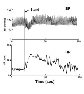 Immediate Orthostatic Hypotension (IOH) upon standing. There is a short-lived decrease in blood pressure (BP - upper panel) and increase in heart rate (HR - lower panel). The fall in BP is resolved within 20 seconds. The patient experienced transient lightheadedness.
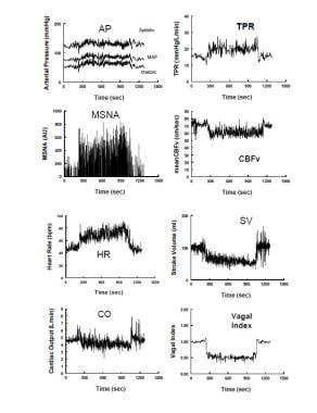 Hemodynamic and neurovascular changes during upright tilt in a representative healthy volunteer. The left panel shows from top to bottom: arterial pressure, muscle sympathetic nerve activity (MSNA) from the peroneal nerve, heart rate (HR) and cardiac output. The right panel shows from top to bottom: total peripheral resistance (TPR), cerebral blood flow velocity (CBFv) by transcranial Doppler ultrasound, stroke volume and a vagal index calculated from the respiratory sinus arrhythmia component of the frequency spectrum of HR variability. During upright tilt at 275 seconds (s), systolic, diastolic and arterial pressures increase slightly, while pulse pressure is decreased with a decrease in stroke volume by approximately 40%. HR increases so that cardiac output is only decreased by 20% because of the increase in HR. CBFv decreases by 5-10%. Both total peripheral vascular resistance and muscle sympathetic nerve activity increase, while the vagal index decreases, reflecting, respectively, sympathetic activation and parasympathetic withdrawal.
