Background
Herpes simplex viruses are ubiquitous, host-adapted pathogens that cause a wide variety of disease states. Two types exist: herpes simplex virus type 1 (HSV-1) and type 2 (HSV-2). Both are closely related but differ in epidemiology. HSV-1 is traditionally associated with orofacial disease, whereas HSV-2 is traditionally associated with genital disease. Lesion location, however, is not necessarily indicative of viral type, as HSV-1 is associated with genital infections more often than HSV-2 in some unique subpopulations.
The term herpes is derived from the Greek word “to creep or crawl” and dates back to early Greek civilization, approximately 2000 years ago, in reference to the spreading nature of herpetic skin lesions.
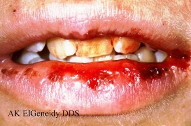 Herpes simplex virus type 1. Primary herpes can affect the lips, and the ruptured vesicles may appear as bleeding of the lips. Courtesy of A.K. ElGeneidy, DDS.
See Herpes Simplex Viruses: Test Your Knowledge, a Critical Images slideshow, for more information on clinical, histologic, and radiographic imaging findings in HSV-1 and HSV-2.
Also, see the 20 Signs of Sexually Transmitted Infections and Clues in the Oral Cavity: Are You Missing the Diagnosis? slideshows to help make an accurate diagnosis.
Up to 80% of herpes simplex infections are asymptomatic. Symptomatic infections can be characterized by significant morbidity and recurrence. In immunocompromised hosts, infections can cause life-threatening complications.
The prevalence of HSV infection worldwide has increased over the last several decades, making it a major public health concern. Prompt recognition of herpes simplex infection and early initiation of therapy are of utmost importance in the management of the disease.
Microbiology
HSV belongs to the alpha herpesvirus group. It is an enveloped virus that is approximately 160 nm in diameter with a linear, double-stranded DNA genome. The overall sequence homology between HSV-1 and HSV-2 is about 50%. HSV-1 has tropism for oral epithelium, while HSV-2 has tropism for genital epithelium. HSV infection is mediated through attachment via ubiquitous receptors to cells, including sensory neurons, leading to establishment of latency. [1]
Pathophysiology
HSV-1 and HSV-2 are characterized by the following unique biological properties [1] :
-
Neurovirulence (the capacity to invade and replicate in the nervous system)
-
Latency (the establishment and maintenance of latent infection in nerve cell ganglia proximal to the site of infection): In orofacial HSV infections, the trigeminal ganglia are most commonly involved, while, in genital HSV infection, the sacral nerve root ganglia (S2-S5) are involved.
-
Reactivation: The reactivation and replication of latent HSV, always in the area supplied by the ganglia in which latency was established, can be induced by various stimuli (eg, fever, trauma, emotional stress, sunlight, menstruation), resulting in overt or covert recurrent infection and shedding of HSV. In immunocompetent persons who are at an equal risk of acquiring HSV-1 and HSV-2 both orally and genitally, HSV-1 reactivates more frequently in the oral rather than the genital region. On the other hand, HSV-2 reactivates 8-10 times more commonly in the genital region than in the orolabial regions. Reactivation is more common and severe in immunocompromised individuals. [2]
Cellular immunity is an important defense against herpes simplex. Dissemination of herpes simplex infection can occur in people with impaired T-cell immunity, such as in organ transplant recipients and in individuals with AIDS. Herpes simplex infection can also complicate burn wounds or damaged skin such as in atopic dermatitis or other allergic dermatoses.
HSV is distributed worldwide. Humans are the only natural reservoirs, and no vectors are involved in transmission. Endemicity is easily maintained in most human communities owing to latent infection, periodic reactivation, and asymptomatic virus shedding. [3]
HSV is transmitted by close personal contact, and infection occurs via inoculation of virus into susceptible mucosal surfaces (eg, oropharynx, cervix, conjunctiva) or through small cracks in the skin. The virus is readily inactivated at room temperature and by drying; hence, aerosol and fomitic spread are rare.
Epidemiology
Frequency
United States
HSV is the most common cause of genital ulcers in the United States. HSV-1 is usually acquired in childhood by contact with oral secretions that contain the virus. The presence of HSV-2 can be used as an indirect measure of sexual activity. Seroprevalence rates do not reflect how many of these individuals have or will have symptomatic episodes of HSV recurrence, as the presence of antibodies is poorly correlated with disease protection. Epidemiology of HSV-1 infection in the US is undergoing a remarkable and subtle transition, with less exposure in childhood and more in adulthood, and less oral acquisition but more genital acquisition. [4] HSV-1 could be overtaking HSV-2 as the main cause of first episode of genital herpes in the United States and elsewhere. [5, 6] In a study of college students in the US, the percentage of genital herpes attributed to HSV-1 (as opposed to HSV-2) increased from 31% in 1993 to 78% in 2001. [6]
Seroprevalence:
Based on the National Health and Nutrition Examination Survey (NHANES) during 2015–2016, prevalence of herpes simplex virus type 1 (HSV-1) was 47.8%, and prevalence of herpes simplex virus type 2 (HSV-2) was 11.9%. Prevalence of both HSV-1 and HSV-2 increased with age. Antibodies to HSV-1 increase with age starting in childhood and correlate with socioeconomic status, race, and cultural group. By age 30 years, 50% of individuals in a high socioeconomic status and 80% in a lower socioeconomic status are seropositive. Antibodies to HSV-2 begin to emerge at puberty, correlating with the degree of sexual activity. More than 90% of adults have antibodies to HSV-1 by the fifth decade of life. [1] A slight crossover of immunity occurs between HSV-1 and HSV-2, allowing for milder subsequent infection by the partner virus type.
International
HSV is well distributed worldwide, with over 23 million new cases per year. An increase in seroprevalence of antibodies to HSV-2 has been documented throughout the world (including the United States) over the last 20 years. [1]
Mortality/Morbidity
Morbidity and mortality rates associated with HSV infections are discussed in Complications. Overall, the mortality rate associated with herpes simplex infections is related to 3 situations: perinatal infection, encephalitis, and infection in the immunocompromised host.
Race
HSV-2 is most prevalent among non-Hispanic blacks (40.3%) compared with the members of other US racial/ethnic groups; 13.7% among non-Hispanic whites and 11.9% among Mexican Americans. [7]
Sex
Seropositivity to HSV-2 is more common in women (25%) than in men (17%). [8]
Age
HSV-1 infections transmitted via saliva are common in children, although primary herpes gingivostomatitis can be observed at any age. HSV-2 infections are clustered perinatally (from a maternal episode at delivery) and primarily once sexual activity begins. HSV-2 genital infections in children can be an indication of sexual abuse. Increased age (after onset of sexual activity) and total number of sexual partners are independent factors associated with increased seroprevalence of HSV-2 antibodies. [8]
-
Herpes simplex virus type 1. Primary herpes can affect the lips, and the ruptured vesicles may appear as bleeding of the lips. Courtesy of A.K. ElGeneidy, DDS.
-
Herpes simplex virus type 1. Recurrent herpes is most often noted clinically as herpes labialis, with clustered vesicles (often coalescing) on the lip vermilion and often on the perioral skin. Recurrences generally occur in the same area each time, although their severity may vary. Courtesy of Sara Gordon, DDS.
-
This neonate displayed a maculopapular outbreak on his feet due to congenitally acquired herpes simplex virus infection. Courtesy of the CDC/Judith Faulk.
-
Herpes simplex virus type 1. Recurrent herpes is occasionally observed intraorally. Inside the oral cavity, recurrent herpes typically affects only keratinized tissues, such as the gingiva or the hard palate. Vesicles often break quickly, so the clinician may observe small clustered ulcers. Courtesy of Sheldon Mintz, DDS.
-
Herpetic whitlow in a young child who earlier had developed herpes gingivostomatitis. Courtesy of Wikimedia Commons [James Heilman, MD] (https://commons.wikimedia.org/w/index.php?search=Herpetic+whitlow+in+a+young+child&title=Special:MediaSearch&go=Go&type=image).
-
Eczema herpeticum; cluster of blisters and punched out erosions of HSV in a child. Courtesy of Wikimedia Commons [Mohammad2018] (https://commons.wikimedia.org/wiki/File:Eczema_herpitcum.jpg).
-
HSV encephalitis; coronal T2-weighted MRI showing increased intensity in the temporal lobes (arrow) in a 33-year-old female who presented with fever, confusion, agitation, and mutism and was diagnosed with HSV encephalitis. Courtesy of Wikimedia Commons [Dr Laughlin Dawes] (https://commons.wikimedia.org/wiki/File:Hsv_encephalitis.jpg).
-
Genital herpes (female); outbreak of genital herpes affecting the vulva. Courtesy of Wikimedia Commons [SOA-AIDS Amsterdam] (https://commons.wikimedia.org/wiki/File:SOA-Herpes-genitalis-female.jpg).
-
Genital herpes (male); vesicular lesions of HSV affecting the penis. Courtesy of Wikimedia Commons [SOA-AIDS Amsterdam] (https://commons.wikimedia.org/wiki/File:SOA-Herpes-genitalis-male.jpg).
Tables

What would you like to print?
- GSK's Experimental Herpes Vaccine Fails to Meet Main Goal in Trial
- Oral Herpes Tied to Double Dementia Risk in Older Adults
- Traumatic Brain Injury May Reactivate Herpes Virus Leading to Neurodegeneration
- Acute Liver Failure due to Herpes Simplex Viral Hepatitis Diagnosed by Skin Lesions and Blood Tests
-
 Epstein-Barr and MS: Just How Strong Is the Link?
-
USPSTF Recommends Against Routine Herpes Screening for Asymptomatic Teens and Adults