Overview
The brachial plexus (plexus brachialis) is a somatic nerve plexus formed by intercommunications among the ventral rami (roots) of the lower four cervical nerves (C5-C8) and the first thoracic nerve (T1). The plexus, depicted in the images below, is responsible for the motor innervation of all the muscles of the upper extremity, with the exception of the trapezius (spinal accessory nerve) and levator scapula (third and fourth cervical nerves and dorsal scapular nerve). [1, 2]
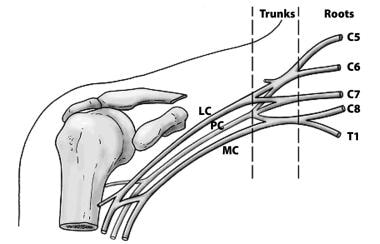 The basic anatomical relationships of the brachial plexus (BP). The BP is subdivided into roots, trunks, divisions, cords, and branches. LC stands for lateral cord, PC stands for the posterior cord, and MC stands for the medial cord.
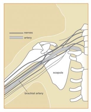 Diagram showing relationships of the brachial plexus (BP) to the sternum, scapula, and humerus.
The brachial plexus supplies all the cutaneous innervation of the upper limb, except for the area of the axilla (which is supplied by the supraclavicular nerve) and the dorsal scapula area, which is supplied by the cutaneous branches of the dorsal rami.
The brachial plexus communicates with the sympathetic trunk via the gray rami communicantes, which join the roots of the plexus. They are derived from the middle and inferior cervical sympathetic ganglia and the first thoracic sympathetic ganglion.
Gross Anatomy
Brachial plexus architecture
The brachial plexus is subdivided into roots, trunks, divisions, cords, and branches. Several mnemonics can be used to remember this architecture (e.g., Really Tired Drink Coffee Black). Typically, the brachial plexus is composed of five roots, three trunks, six divisions, three cords, and terminal branches, as seen in the image below.
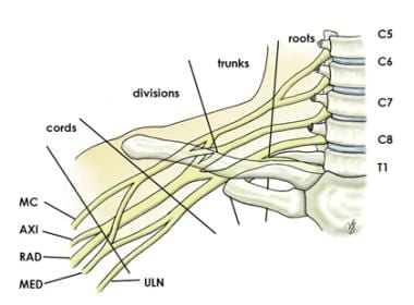 Brachial plexus with terminal branches labeled. MC is musculocutaneous (nerve), AXI is axillary, RAD is radial, MED is median, and ULN is ulnar.
Roots
The ventral rami of spinal nerves C5 to T1 are referred to as the "roots" of the plexus. The typical spinal nerve root results from the confluence of the ventral nerve rootlets originating in the anterior horn cells of the spinal cord and the dorsal nerve rootlets that join the spinal ganglion in the region of the intervertebral foramen.
The roots emerge from the transverse processes of the cervical vertebrae immediately posterior to the vertebral artery, which travels in a cephalocaudad direction through the transverse foramina. Each transverse process consists of a posterior and anterior tubercle, which meet laterally to form a costotransverse bar. The transverse foramen lies medial to the costotransverse bar and between the posterior and anterior tubercles. The spinal nerves that form the brachial plexus run in an inferior and anterior direction within the sulci formed by these structures.
Trunks
Shortly after emerging from the intervertebral foramina, the five roots (C5-T1) unite to form three trunks. The trunks of the brachial plexus pass between the anterior and middle scalene muscles.
The ventral rami of C5 and C6 unite to form the upper trunk. The suprascapular nerve and the nerve to the subclavius arise from the upper trunk. The suprascapular nerve contributes sensory fibers to the shoulder joint and provides motor innervation to the supraspinatus and infraspinatus muscles.
The ventral ramus of C7 continues as the middle trunk. The ventral rami of C8 and T1 unite to form the lower trunk.
Divisions
Each trunk splits into an anterior division and a posterior division. These separate the innervation of the ventral and dorsal aspect of the upper limb. The anterior divisions usually supply flexor muscles.
These include muscles such as the biceps brachii, brachialis, and coracobrachialis. [2, 3] The posterior divisions usually supply extensor muscles such as the triceps brachii, brachioradialis, and latissimus dorsi. This division is consistent across the upper limb, helping to control coordinated movement and force distribution between flexion and extension. [2, 3]
Cords
The cords are referred to as the lateral, posterior, and medial cord, according to their relationship with the axillary artery, as seen in the image below. The cords pass over the first rib close to the dome of the lung and continue under the clavicle immediately posterior to the subclavian artery.
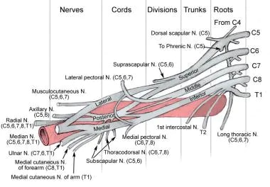 Diagram showing basic relationships of the brachial plexus to the pectoralis minor muscle and the axillary artery, which is a continuation of the subclavian artery.
The anterior divisions of the upper and middle trunks unite to form the lateral cord, which is the origin of the lateral pectoral nerve (C5, C6, and C7). This nerve supplies the pectoralis major muscle. [2, 3, 4]
The anterior division of the lower trunk forms the medial cord, which gives off the medial pectoral nerve (C8, T1), the medial brachial cutaneous nerve (T1), and the medial antebrachial cutaneous nerve (C8, T1), which provide sensory innervation to the skin of the arm and forearm, respectively. [2, 3, 4] The posterior divisions from each of the three trunks unite to form the posterior cord.
The upper (C7, C8) and lower (C5, C6) subscapular nerves leave the posterior cord and descend behind the axillary artery to supply the subscapularis and teres major muscles. The thoracodorsal nerve to the latissimus dorsi (also known as the middle subscapular nerve [C6, C7, and C8]) also arises from the posterior cord, as seen in the image below.
These cords continue to give rise to major terminal branches of the brachial plexus such as the radial, axillary, median, ulnar, and musculocutaneous nerves, which innervate various muscles of the upper limb and provide sensory functions. [2, 3, 4]
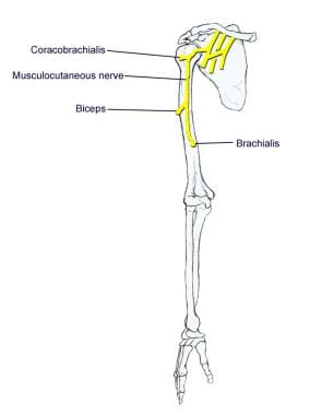
Musculocutaneous nerve branch
The musculocutaneous nerve is a mixed nerve that contains sensory and motor axons. The musculocutaneous nerve is derived from the lateral cord. The musculocutaneous nerve originates from the lateral cord of the brachial plexus, typically arising from spinal nerves C5 to C7. [5] The musculocutaneous nerve leaves the brachial plexus sheath high in the axilla at the level of the lower border of the teres major muscle and passes into the coracobrachialis muscle. It innervates the muscles in the flexor compartment of the arm and carries sensation from the lateral (radial) side of the forearm. (See the image below.) In terms of distance measurements, the average distance from the coracoid process to where the musculocutaneous nerve (MCN) enters the coracobrachialis is approximately 5.6 cm. MCN is primarily responsible for innervating the flexor compartment of the arm, which includes the biceps brachii and brachialis muscles, and it also provides sensory innervation to the lateral aspect of the forearm via the lateral cutaneous nerve of the forearm. [5]
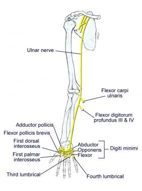
Ulnar nerve branch
The ulnar nerve is derived from the medial cord, specifically from spinal roots C8 and T1. It travels distally through the axilla, arm, and forearm into the hand. [5, 6] Motor innervation is mainly to the intrinsic muscles of the hand (as seen in the image below). Sensory innervation is to the medial (ulnar) 1.5 digits (little finger, half of the ring finger).
Motor innervation: The ulnar nerve primarily provides motor innervation to: [5, 6]
-
Flexor carpi ulnaris - This muscle aids in flexion and adduction of the wrist
-
Flexor digitorum profundus (medial half) - Responsible for flexing the ring and little fingers at the distal interphalangeal joints
In addition to these forearm muscles, the ulnar nerve innervates most of the intrinsic muscles of the hand, excluding those supplied by the median nerve. Key muscles include: [5, 6]
-
Hypothenar muscles - These include the flexor digiti minimi brevis, abductor digiti minimi, and opponens digiti minimi
-
Interossei muscles - Both the palmar and dorsal interossei are innervated by the ulnar nerve
-
Lumbricals - The two medial lumbricals receive motor supply from the ulnar nerve
-
Adductor pollicis - This muscle is crucial for thumb adduction
The ulnar nerve primarily controls the intrinsic muscles of the hand, excluding the thenar muscles and the two lateral lumbricals. The superficial branch of the ulnar nerve innervates the palmaris brevis muscle, while its deep branch supplies the majority of intrinsic hand muscles. [5, 6]
Sensory innervation: The sensory distribution of the ulnar nerve covers: [5, 6]
-
The medial one and a half digits (the little finger and half of the ring finger)
-
The associated palm area through its palmar cutaneous branch, which innervates the medial half of the palm
-
The dorsal surface of the same medial one and a half fingers via its dorsal cutaneous branch, which emerges in the forearm before entering the hand
Injuries to the ulnar nerve, particularly at the elbow (e.g., cubital tunnel syndrome) or wrist (e.g., Guyon canal syndrome), can lead to motor and sensory deficits such as ulnar claw hand and loss of finger movement. [5, 6]
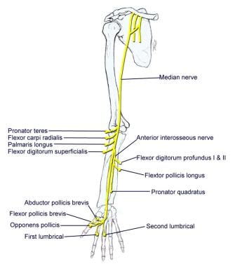
Median nerve branch
The median nerve is derived from the lateral and medial cords with contributions from the spinal roots C6-C7 (lateral) and C8-T1 (medial). [7] Motor innervation is to most flexor muscles in the forearm and intrinsic muscles of the thumb (thenar muscles), as seen in the image below. Sensory innervation is to the lateral (radial) 3.5 digits (thumb, index, and middle fingers; half of the ring finger).
Motor innervation: The median nerve innervates most of the flexor muscles in the forearm and several intrinsic muscles of the hand, particularly those involved in thumb movement. The specific muscles innervated by the median nerve include: [7, 8, 9]
-
Superficial layer - Pronator teres, flexor carpi radialis, palmaris longus, flexor digitorum superficialis
-
Deep layer - Flexor pollicis longus, pronator quadratus, and the lateral half of flexor digitorum profundus (the medial half is innervated by the ulnar nerve)
-
Thenar muscles - Abductor pollicis brevis, opponens pollicis, and the superficial head of the flexor pollicis brevis via the recurrent branch
-
Lateral lumbricals - Innervated by the palmar digital branches
This distribution allows for essential functions such as wrist flexion and finger movements, including opposition and abduction of the thumb.
Sensory innervation: The median nerve provides sensory innervation to the palmar aspect of the hand, specifically: [7, 8, 9]
-
Lateral aspect of the palm
-
Palmar surface and fingertips of the thumb, index finger, middle finger, and half of the ring finger
The palmar cutaneous branch, which arises before the median nerve enters the carpal tunnel, supplies sensation to a portion of the palm but does not pass through the carpal tunnel itself.
As it courses through the forearm, the median nerve travels between the two heads of pronator teres and descends between the flexor digitorum superficialis and flexor digitorum profundus. It gives rise to two major branches: [7, 8, 9]
-
Anterior interosseous nerve - Supplies deep flexor muscles
-
Palmar cutaneous nerve - Provides sensory innervation to part of the palm
Upon reaching the wrist, it enters the hand through the carpal tunnel alongside tendons of the flexor digitorum superficialis and flexor digitorum profundus. The median nerve typically bifurcates into a recurrent branch (for thenar muscles) and digital branches for sensory innervation. [7, 8, 9]
This nerve can be involved in conditions such as the carpal tunnel syndrome, where compression leads to numbness, tingling, and weakness in these digits, particularly affecting the thumb function. Another condition is pronator teres syndrome, where compression of the nerve in the forearm causes pain and weakness in the forearm and hand. [7, 8, 9]
Axillary nerve branch
The axillary nerve is derived from the posterior cord, with fibers from C5 and C6. [10] The axillary nerve leaves the brachial plexus at the lower border of the subscapularis muscle and continues along the inferior and posterior surface of the axillary artery as the radial nerve.
It typically exits the brachial plexus at the lower border of the subscapularis muscle, traveling closely along the inferior and posterior surfaces of the axillary artery. As it courses through the quadrangular space, it divides into anterior and posterior branches, providing vital motor and sensory functions. [10]
Motor innervation: The axillary nerve serves as motor innervation to the deltoid and teres minor muscles, as seen in the image below. These act at the glenohumeral joint: [10]
-
Deltoid muscle - Responsible for shoulder abduction, flexion, and extension
-
Teres minor muscle - Aids in external rotation of the arm
Sensory innervation:
Sensory innervation is from the skin just below the point of the shoulder. The axillary nerve continues as the superior lateral brachial cutaneous nerve of the arm.
The nerve is closely associated with the posterior circumflex humeral artery and vein as it passes through the quadrangular space. The axillary nerve can be injured by shoulder dislocations, humeral fractures, or the improper use of crutches, leading to muscle paralysis and sensory loss in the affected area. Recovery from such injuries can be slow and may require physical therapy or surgical intervention in severe cases. [10]
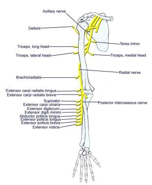
Radial nerve branch
The radial nerve is also derived from the posterior cord (nerve roots C5 to T1). [2] The radial nerve continues along the posterior and inferior surface of the axillary artery and innervates the extensor muscles of the elbow, wrist, and fingers, as seen in the image above. As it descends along the posterior aspect of the humerus, it travels within a shallow groove known as the radial groove. This nerve wraps around the humerus laterally and continues down toward the elbow, where it enters the forearm by passing anterior to the lateral epicondyle through the cubital fossa. Upon reaching the forearm, the radial nerve bifurcates into two main branches: [11, 12]
-
Deep branch (motor) - This branch innervates muscles in the posterior compartment of the forearm
-
Superficial branch (sensory) - This branch provides sensory innervation to the skin on the dorsum of the hand and fingers, particularly affecting the dorsal surface of the lateral three and a half digits
Motor functions: The radial nerve is responsible for innervating several key muscles: [11, 12]
-
Triceps brachii - Responsible for elbow extension
-
Brachioradialis - Assists in flexing the elbow
-
Several other extensor muscles - Includes the extensor carpi radialis longus and brevis, extensor digitorum, and extensor carpi ulnaris, which facilitate wrist and finger extension.
The deep branch specifically innervates muscles such as: [11, 12]
-
Supinator
-
Extensor carpi radialis brevis
-
The remaining deep extensors of the forearm
Sensory functions: The sensory distribution of the radial nerve includes: [11, 12]
-
Posterior cutaneous nerve of arm - Innervates the skin on the posterior surface of the arm
-
Posterior cutaneous nerve of the forearm - Supplies sensation to a strip down the middle of the posterior forearm
-
Superficial branch - Provides cutaneous sensation to the dorsal aspect of fingers one through three and part of the ring finger
Compression of the radial nerve can occur at several sites, including the arcade of Frohse and the radial tunnel, leading to syndromes such as radial tunnel syndrome or posterior interosseous nerve syndrome. [11, 12]
Additional branches
In addition to the five terminal branches described above, numerous preterminal or collateral branches leave the plexus at various points along its length.
Dorsal scapular nerve
The dorsal scapular nerve is derived from the C5 root just after its exit from the intervertebral foramen. It serves as the motor nerve to the rhomboids major and minor muscles which are essential for scapular retraction and stabilization. [2, 3]
Long thoracic nerve
The long thoracic nerve is derived from C5, C6, and C7 roots immediately after their emergence from the intervertebral foramina. The long thoracic nerve crosses the first rib and then descends through the axilla behind the major branches of the plexus. It innervates the serratus anterior muscle, which is vital for scapular protraction and upward rotation. It is important in maintaining shoulder stability and function. It is prone to injuries, leading to isolated long thoracic nerve palsy, which can lead to scapular winging. [2, 3]
Phrenic nerve
The phrenic nerve arises from the C3, C4, and C5 root levels, although chiefly from the C4 nerve root. It crosses the anterior scalene from lateral to medial and extends into the thorax between the subclavian vein and artery. This nerve is critical because it is majorly responsible for diaphragm movement, thereby playing a key role in respiration. [2, 3]
Subclavius muscle nerve
The nerve to the subclavius muscle is a small filament that arises from the upper trunk (C5-C6). It descends to the subclavius muscle (which helps stabilize the clavicle and shoulder gridle) in front of the subclavian artery and the lower trunk of the plexus. [2]
Suprascapular nerve
The suprascapular nerve arises from the upper trunk formed by the union of the fifth and sixth cervical nerves. It innervates the supraspinatus muscles and infraspinatus muscles. It runs laterally beneath the trapezius and the omohyoideus and enters the supraspinatus fossa through the suprascapular notch, below the superior transverse scapular ligament; it then passes beneath the supraspinatus and curves around the lateral border of the spine of the scapula to the infraspinatus fossa.
Lateral pectoral nerve
The lateral pectoral nerve arises from the lateral cord of the brachial plexus, from the fifth, sixth, and seventh cervical nerves. It passes across the axillary artery and vein, pierces the coracoclavicular fascia, and is distributed to the deep surface of the pectoralis major. It sends a filament to join the medial anterior thoracic and forms a loop with it in front of the first part of the axillary artery. This nerve innervates the clavicular head of the pectoralis major muscle.
Medial pectoral nerve
The medial pectoral nerve arises from the medial cord from the eighth cervical and first thoracic nerve. It passes behind the first part of the axillary artery, curves forward between the axillary artery and vein, and unites in front of the artery with a filament from the lateral nerve. It then enters the deep surface of the pectoralis minor, where it divides into a number of branches, which supply the muscle. Several branches of the medial pectoral nerve pierce the muscle and end in the pectoralis major, which supply the muscle.
The medial and lateral pectoral nerve often join together to act as a single nerve innervating the pectoralis major and minor muscles.
Medial brachial cutaneous nerve
The medial brachial cutaneous nerve is the smallest branch of the brachial plexus; arising from the medial cord, it receives its fibers from the eighth cervical and first thoracic nerves. It passes through the axilla, at first lying behind and then medial to the axillary vein and communicates with the intercostobrachial nerve.
The medial brachial cutaneous nerve descends along the medial side of the brachial artery to the middle of the arm, where it pierces the deep fascia, and is distributed to the skin of the back of the lower third of the arm, extending as far as the elbow, where some filaments are lost in the skin in front of the medial epicondyle and others over the olecranon. It communicates with the ulnar branch of the medial antebrachial cutaneous nerve. The medial brachial cutaneous nerve carries sensation from the lower medial portion of the arm.
Medial antebrachial cutaneous nerve
The medial antebrachial cutaneous nerve arises from the medial cord of the brachial plexus. It derives its fibers from the eighth cervical and first thoracic nerves and at its commencement is medial to the axillary artery. It gives off a filament near the axilla that pierces the fascia and supplies the integument covering the biceps brachii, nearly as far as the elbow. The nerve then runs down the ulnar side of the arm medial to the brachial artery, pierces the deep fascia with the basilic vein, about the middle of the arm, and divides into a volar and an ulnar branch.
Blood Supply of the Brachial Plexus
The blood supply of the brachial plexus is based largely on the subclavian (which becomes the axillary) artery and its branches, and variations exist. Generally, the vessels involved are the vertebral, ascending and deep cervical, and superior intercostal arteries. The cord and rootlets of the cervical nerves are supplied by the anterior and posterior spinal branches of the vertebral artery. The trunks of the plexus are supplied by muscular branches of the ascending and deep cervical arteries and superior intercostals and occasionally by the subclavian itself. [13]
Natural Variants
Many variant forms of the brachial plexus exist, with none representing most patients. Kerr catalogued 29 forms of the brachial plexus in some 175 cadaver specimens dissected between 1895 and 1910. In the early part of the last century, one author described a total of 38 variations of the plexus. Up to 53.5% of plexuses in cadaver studies possess significant anatomic variation from the "classic" description of the brachial plexus.
A study published in September 2003 found variants in 107 of 200 fetuses examined. The authors pointed out that morphologic variations were more common in female fetuses and were found most often on the right side. [14]
A prefixed brachial plexus (cephalic or high) occurs when the C4 ventral ramus contributes to the brachial plexus; contributions to the plexus usually come from the C4-C8. A postfixed brachial plexus (caudal or low) occurs when the T2 ventral ramus contributes to the brachial plexus; contributions to the plexus usually come from C6-T2.
A 2023 study identified 17.8% of 140 specimens displaying variations in the origins and segments of the brachial plexus, with a notable prevalence of prefixed configurations (12 variations on the right side and eight on the left) and some postfixed configurations: [15, 16, 17, 18, 19, 20]
-
Prefixed brachial plexus - Occurs when contributions include the C4 ventral ramus alongside C5-C8
-
Postfixed brachial plexus - Involves contributions from T2 along with C6-T1
These configurations can significantly affect surgical approaches and outcomes, necessitating a thorough understanding by healthcare professionals involved in upper limb surgeries. [15, 16, 17, 18, 19, 20]
A meta-analysis published in 2022 highlighted that variations are common and can lead to significant clinical implications, including altered nerve territories and potential complications during surgical interventions. [15, 16, 17, 18, 19, 20]
Another study found that 56% of the examined plexuses had typical origins, while 9.4% were prefixed and 3.1% were postfixed, indicating a substantial occurrence of atypical configurations. [15, 16, 17, 18, 19, 20]
The clinical implications of these variations are profound. Variations may lead to conditions such as neuralgias or altered motor innervation patterns in the upper limb. Some studies have specifically pointed out that nerve branching variations can be associated with thoracic outlet syndrome symptoms, particularly when nerves pierce through scalene muscles. [15, 16, 17, 18, 19, 20]
Lesions of the Brachial Plexus
Knowledge of the muscles innervated by branches of the brachial plexus and the actions of these muscles and areas of anesthesia and/or paresthesia allows the physician to determine the localization of a given lesion.
Brachial plexus injuries have numerous causes such as labor and delivery. A plexus injury at birth is likely caused by a stretch or tear of the child's brachial plexus during the delivery process. The abducted arm of the infant can get pinned against the child's head. This injury results in incomplete sensory and/or motor function of the injured nerve. Traumatic brachial plexus injuries may occur due to motor vehicle accidents, bike accidents, all-terrain vehicle accidents, or sports.
Nerve injuries vary in severity from a mild stretch to the nerve root tearing away from the spinal cord and include the following:
-
Avulsion - The nerve is torn away from its attachment at the spinal cord; this is the most severe type of injury
-
Rupture - The nerve is torn but not at the spinal cord attachment
-
Neuroma - Scar tissue has grown around the injury site, putting pressure on the injured nerve and preventing the nerve from sending signals to the muscles
-
Neurapraxia - The nerve has been stretched and damaged but not torn
Diagnoses Related to Brachial Plexus Injuries
Diagnoses related to brachial plexus injuries include Erb's palsy, Klumpke's palsy, thoracic outlet syndrome, Burner syndrome, and Parsonage-Turner syndrome.
Erb's palsy
Erb's palsy involves the upper root nerves of C5, C6. The patient often presents with the arm extended and wrist fully flexed (waiter's tip).
Motor deficits are loss of abduction, flexion, and rotation at the shoulder (axillary, suprascapular, upper, and lower subscapular nerve); weak shoulder extension; and weak elbow flexion and supination of the radioulnar joint (musculocutaneous and radial nerve). The patient is susceptible to shoulder dislocation due to loss of rotator cuff muscles.
Sensory deficits are to the posterior and lateral aspect of the arm (axillary nerve), radial side of the forearm (musculocutaneous nerve), and thumb and first finger (superficial branch of the radial nerve; digital branches of the median nerve).
Studies emphasize the importance of timely intervention for optimal recovery outcomes. Surgical options may include nerve grafting or transfers, with better prognosis when performed within 6 months post-injury. [21, 22, 23]
Klumpke's palsy
This condition involves the lower root nerves of C8 and T1. Klumpke's palsy is caused by a rare injury of the lower brachial plexus, usually following breech delivery.
Motor deficits are opposite to the thumb (thenar branch of the median nerve), loss of adduction of the thumb (ulnar nerve), loss of abduction and adduction of metacarpophalangeal joints, flexion at the metacarpophalangeal and extension of the interphalangeal joints (deep branch of the ulnar and median nerve), and weak flexion of the proximal interphalangeal joints and distal interphalangeal joints (ulnar and median nerve).
Sensory deficit is to the ulnar side of the forearm, hand, and ulnar 1.5 digits (ulnar and medial antebrachial cutaneous nerve).
Symptoms include paralysis or weakness in the hand and forearm, with characteristic "claw hand" deformity. In some cases, it can lead to Horner syndrome, involving ptosis, miosis, and anhidrosis. [21, 22, 23]
Research indicates that early diagnosis and intervention can significantly improve functional outcomes in infants with Klumpke's palsy. [21, 22, 23]
Thoracic outlet syndrome
Thoracic outlet syndrome (TOS) is caused by compression of the neurovascular structures (subclavian vessels, brachial plexus, cervical ganglia, and vertebral artery) in the cervicoaxillary region. It may be congenital or acquired. Symptoms vary widely but often include arm pain, tingling, or weakness in the arm. Vascular symptoms include swelling and discoloration. [21, 22, 23]
Bony causes include the long transverse process of the seventh cervical vertebra, cervical rib, anomalous first rib, clavicle fracture, and first rib fracture. Soft tissue causes include congenital bands, poor posture, mass lesions, cervical strain, hypertrophy, and/or injury of the anterior and middle scalene muscles or the pectoralis minor muscle.
Surgical approaches have shown promise in managing venous TOS effectively. Techniques such as first rib resection have demonstrated durable outcomes for patients with axillary-subclavian vein thrombosis due to TOS. [21, 22, 23]
Burner syndrome
This condition is believed to be related to brachial plexus stretch and/or nerve root compression. It generally involves the upper trunk of the brachial plexus, particularly C5-C6. Burner syndrome is usually a result of traction injury, with axial distraction or downward force on the shoulder combined with lateral bend of the neck away from the shoulder.
Brachial plexus stretch injuries are found in young athletes, with neck pain being less common. Nerve root compression is found in older athletes (college) and is generally associated with cervical disc disease or cervical stenosis. Neck pain is more common in older athletes.
Parsonage-Turner syndrome (brachial neuritis)
This condition is generally associated with a viral prodrome, immunizations, and significant pain. Acute, excruciating, unilateral shoulder pain is present, followed by flaccid paralysis of the shoulder and parascapular muscles several days later.
Parsonage-Turner syndrome varies greatly in manifestation and nerve involvement. Due to the extreme pain involved, patients usually present acutely. Often, the affected arm is supported by the uninvolved arm and is held in adduction and internal rotation.
The patient may have atrophy of the affected muscles. Pain may occur with palpation and active and passive range of motion. Reflexes may be reduced or absent depending on the nerves involved. Sensory loss is usually not prominent, although it may be detectable. Parsonage-Turner syndrome may cause scapular winging.
Symptoms often include severe unilateral shoulder pain followed by muscle weakness. Flaccid paralysis affects the shoulder and scapular muscles. Patients typically present with significant pain before developing weakness days later. Management focuses on pain control and rehabilitation. Recovery can take months to years, with varying degrees of nerve involvement. [21, 22, 23]
-
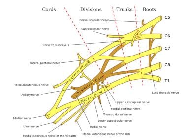
Schema of the brachial plexus.
-
The basic anatomical relationships of the brachial plexus (BP). The BP is subdivided into roots, trunks, divisions, cords, and branches. LC stands for lateral cord, PC stands for the posterior cord, and MC stands for the medial cord.
-
Diagram showing basic relationships of the brachial plexus to the pectoralis minor muscle and the axillary artery, which is a continuation of the subclavian artery.
-
Diagram showing relationships of the brachial plexus (BP) to the sternum, scapula, and humerus.
-
Brachial plexus with terminal branches labeled. MC is musculocutaneous (nerve), AXI is axillary, RAD is radial, MED is median, and ULN is ulnar.
-
Musculocutaneous nerve.
-
Axillary and radial nerves.
-
Median nerve.
-
Ulnar nerve.
-
Nervous system. Full body, anterior view.
