Overview
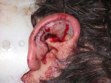
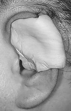
Auricular hematoma, shown below, is a complication that results from direct trauma to the anterior auricle and is a common facial injury in wrestlers. [1, 2, 3] Shearing forces to the anterior auricle can lead to separation of the anterior auricular perichondrium from the underlying, tightly adherent cartilage. This may lead to tearing of the perichondrial blood vessels and subsequent hematoma formation.
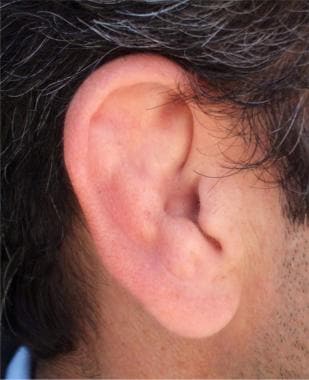
The torn perichondrial vessels compromise the viability of the avascular underlying cartilage. Interestingly, the presence of a subperichondrial hematoma has been found to stimulate new and often asymmetric cartilage to form. [4] This deformity, which is often referred to as cauliflower ear or wrestler’s ear (shown in the image below), is often considered a badge of honor among wrestlers and rugby players. [5]
The goal of treatment is to completely evacuate subperichondrial blood and to prevent its reaccumulation. The mechanism of hematoma drainage has been debated. To date, no randomized controlled trials have addressed this issue. [6]
Relevant anatomy
The auricle and external acoustic meatus (or external auditory canal) compose the external ear. The external ear functions to collect and amplify sound, which then gets transmitted to the middle ear. The asymmetric shape of the external auricle introduces delays in the path of sound that assist in sound localization.
The arterial supply of the auricle is composed of the posterior auricular artery, the anterior auricular branch of the superficial temporal artery, and the occipital artery, which also contributes. Veins accompany the corresponding named arteries.
For more information about the relevant anatomy, see Ear Anatomy.
Indications
Auricular hematoma drainage is indicated for the following:
-
Tender anterior auricular swelling after trauma, which deforms the normal anatomy of the pinna
-
Presentation within 7 days after trauma (After 7 days, the formation of granulation tissue may complicate the procedure. At that point, patients should be referred to a specialist)
Contraindications
Auricular hematoma drainage is contraindicated in the following cases:
-
Hematomas that are older than 7 days
-
Recurrent or chronic hematomas (In such cases, open surgical debridement by a specialist is indicated because the hematoma, granulation tissue, or both can be located within the cartilage instead of in the subperichondrial space.)
Anesthesia
Local anesthesia with lidocaine 1% with or without epinephrine can be infiltrated directly into the area to be incised.
Many authors advocate the use of the lidocaine without the presence of a vasoconstrictive agent such as epinephrine. However, some literature supports the safety of vasoconstrictive agents in areas such as the nose or pinna.
Alternatively, an auricular block can be performed. For more information, see Ear Anesthesia.
Equipment
Equipment needed for auricular hematoma drainage includes:
-
Syringe, 3 mL, with a 23- or 27-gauge (ga) needle for anesthesia
-
Syringe, 10 mL, with a 18- or 20-ga needle (if performing needle aspiration)
-
Lidocaine 1% (with or without epinephrine)
-
Scalpel, No. 15
-
Small suction, if available
-
Irrigation set-up (syringe, normal saline)
-
Compression dressing materials
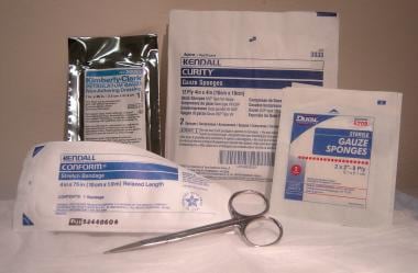
Simple compression dressing, as shown in the image below: dry cotton, Vaseline gauze, 4 x 4 plain gauze, secondary dressing wrap (eg, Kling), scissors
Specialized compression dressing (to be made in a specialist's office; not described here): dental rolls (or cotton bolsters, silicone splints, or plaster mold), nylon or Prolene suture on straight needle
Positioning
Place patient in the lateral decubitus position on the unaffected side.
Technique
Preparation
Cleanse the skin with povidone iodine, ChloraPrep (chlorhexidine gluconate 2% and isopropyl alcohol 70%), or another cleanser.
Anesthetize the area with lidocaine 1% or perform an auricular block. (For more information, see Ear Anesthesia.)
Choose the technique
Technique 1 - Needle aspiration
Although still widely used, this method is no longer recommended by many sources because of hematoma reaccumulation. The aspiration is often inadequate and the hematoma requires additional management. [7] Some sources recommend primary needle aspiration followed by the incision method, if reaccumulation occurs.
Use an 18- or 20-ga needle to aspirate blood from the most fluctuant or full area.
Technique 2 - Incision and drainage
Incise the edge of hematoma along the natural skin folds using a No. 15 scalpel. A small (5 mm) incision is often all that is necessary.
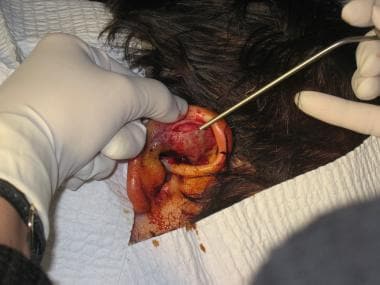
Gently separate the skin and perichondrium from the hematoma and cartilage and completely express or suction out the hematoma, as shown below. Be careful not to damage the perichondrium.
Irrigate the pocket with normal saline with an 18-ga angiocatheter.
Optional step: Leave a small drain in the incision. This allows the wound to drain but also predisposes to infection. If a drain is placed, the patient should always be given antibiotics upon discharge. The drain should be removed in 24 hours if no significant bleeding occurs.
Reapproximate the perichondrium to the cartilage.
Compression dressing
Apply digital pressure for 5-10 minutes, and then apply compression dressing. A simple dressing is inadequate, as the hematoma is likely to reaccumulate.
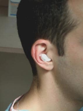
Compression dressing can be applied noninvasively (more applicable in the ED) or surgically. Noninvasive methods include a simple compression dressing or, if available, application of silicone or thermoplastic splints [8] or plaster mold to the medial and lateral aspects of the auricle, as shown below.
Surgical dressing involves securing cotton bolsters, buttons, or thermoplastic splints [9] with through and through sutures to the medial and lateral aspects of the auricle.
A simple compression dressing can be quickly made as follows:
-
Place dry cotton into the external canal, as shown below.
-
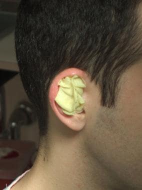
Fill all external auricular crevices with either moist gauze (soaked in saline) or Vaseline gauze, as depicted in the image below.
-
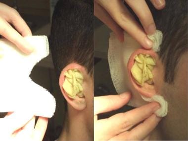
Place 3-4 layers of gauze behind the ear as a posterior gauze pack. Prior to placement, cut out a V-shaped section of gauze so that the gauze fits snugly behind the ear, as shown below.
-
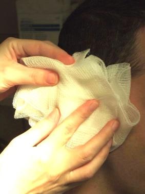
Cover the packed anterior ear as shown below, with multiple layers of fluffed gauze.
-
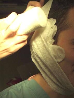
Bandage the fluffed gauze into place with Kling or an elastic bandage, as shown in the image below.
-
Specialized compression dressings, such as a silicone splint or dental rolls sewn onto the anterior and posterior pinna, can also be made, though such dressings are normally prepared and applied by a specialist.
Aftercare
The ear must be reexamined for hematoma reoccurrence every 24 hours for several days.
Aspirin, nonsteroidal anti-inflammatory drugs (NSAIDs), or anticoagulants should be discontinued or avoided for several days to prevent continuing bleeding.
Recommendations indicate that, upon discharge, patients should receive antibiotics that cover common skin flora for 7-10 days. Patients whose immune systems are compromised should receive antibiotic prophylaxis covering both Staphylococcus and Pseudomonas species.
If infections suspicious for Pseudomonas species are discovered during follow-up, the patient should be admitted to the hospital for open drainage and intravenous antibiotics.
Pearls
Do not leave an auricular hematoma undrained unless the injury is older than 7 days.
Apply a compression dressing rather than a simple dressing.
Perform daily follow-up ear examinations.
Complications
Complications may include the following:
-
Site infection
-
Chondritis
-
Scar formation (cauliflower ear) [4]
-
Auricular hematoma.
-
Cauliflower ear.
-
Supplies needed to make a simple compression dressing.
-
Auricular hematoma incision and drainage.
-
Ear splint.
-
Compression dressing: Dry cotton in external canal.
-
Compression dressing: Vaseline gauze in anterior pinna.
-
Compression dressing: Gauze behind pinna.
-
Compression dressing: Gauze applied to anterior ear.
-
Compression dressing: Bandaging dressing into place.
Tables

What would you like to print?
- Otolaryngologic Manifestations of Granulomatosis With Polyangiitis
- Pierre Robin Syndrome
- Skill Checkup: Peritonsillar Abscess Drainage
- Biologic Therapies in Refractory Chronic Rhinosinusitis With Nasal Polyps
- Trending Clinical Topic: Tinnitus
- Fast Five Quiz: Do You Know Best Practices for Antibiotic Use in Respiratory Tract Infections?
- Low-Dose CT Vs Chest X-Ray for Lung Cancer Surveillance in HNSCC: Is One Better?
- Does Vitamin D Deficiency Worsen Pediatric Obstructive Sleep Apnea?
- Can ctDNA Accurately Diagnose HPV-Positive Oropharyngeal Squamous Cell Carcinoma?
-
 American Academy of Otolaryngology–Head and Neck Surgery (AAO-HNS) 2024 Annual Meeting
-
MSK Pediatrics e-Tumor Boards: Case 2: Multisystem Rosai-Dorfman Disease
-
Managing Common Ear Complaints: An ENT's Advice


 A Lump in the Throat: Thyroid Cancer


