Practice Essentials
Meconium plug syndrome (also termed functional immaturity of the colon, colonic immaturity, small left colon syndrome, and functional colonic obstruction) is a transient disorder of the newborn colon characterized by delayed passage (>24-48 hr) of meconium and intestinal dilatation. The incidence is 1 in 500 live births, and it is associated with Hirschsprung disease in up to 40% of cases and cystic fibrosis in up to 40% of cases. An association has been reported between maternal diabetes and meconium plug syndrome. Differential diagnosis includes Hirschsprung disease, malrotation, meconium disease, and intestinal atresia. Hirschsprung disease occurs in one in every 5000 live births. [1, 2, 3, 4, 5, 6, 7, 8, 9]
(See the image below.)
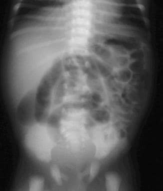 Supine frontal view of the abdomen in a newborn with meconium plug syndrome demonstrates multiple dilated loops of bowel but no rectal gas.
Contrast enema demonstrates the retained meconium as a filling defect or plug that produces a double-contrast effect. Small left colon syndrome is a subset of meconium plug syndrome in which an enema demonstrates an apparent transition zone between the dilated and the normal to decreased caliber distal colon at the splenic flexure. (See the image below.) [3, 4]
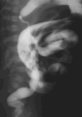 A lateral view from contrast enema in a newborn demonstrates a normal-to-decreased caliber "empty" distal colon and dilated proximal bowel containing multiple plugs. The child responded clinically and radiographically to a single enema.
Preferred examination
The initial imaging modality is plain film radiography, which includes supine and horizontal beam views (left lateral decubitus or cross-table lateral) of the abdomen. Follow plain films with contrast enema. Barium can be used but has been replaced by water-soluble contrast agents in most practices. Historically, Gastrografin was employed, which is a hypertonic solution containing both wetting and detergent agents. However, complications secondary to hyperosmolarity occurred that produced dehydration. Evidence exists that detergent and wetting additives may be toxic, and their possible therapeutic effect remains unproven. [10, 11]
Meconium plug syndrome is a diagnosis of exclusion. Contrast enema usually eliminates congenital small bowel obstruction and rare colon abnormalities (such as atresia and duplication). The primary differential consideration is Hirschsprung disease, which is diagnosed eventually in approximately 10-40% of patients with apparent meconium plug syndrome.
Buonpane et al found that of 373 newborns with meconium plug syndrome (from the Pediatric Health Information System database), 57 (15.3%) were ultimately diagnosed with Hirschsprung disease. However, of 106 of the newborns who underwent rectal biopsy, 49 (40.5%) were diagnosed with Hirschsprung disease. [2]
In a study of 33 very low birthweight infants with meconium obstruction, ultrasound-guided water-soluble contrast enema had an overall success rate of 54.5% (18 of 33 successful cases). Patients in the success group had statistically significant older gestational age, larger birth weight, and higher body weight on the day of the procedure. Retrial of contrast injection during the procedure was associated with significantly higher success than the single trial, and the presence of refluxed contrast into the distal ileum was a statistically significant predictor of success. [5]
Rare disorders that may partially simulate meconium plug syndrome include neuronal intestinal dysplasia, visceral neuropathies, and megacystis-microcolon-intestinal hypoperistalsis syndrome, also termed Berdon syndrome. [12] However, radiographic and clinical features in these diseases usually are distinguished readily from meconium plug syndrome. A more common problem is an infant with sepsis or a metabolic disorder who presents with nonobstructive ileus.
Radiography
Plain films usually demonstrate multiple dilated loops of bowel with absence of rectal gas. The presence or absence of air-fluid levels in the bowel is not helpful. Findings are similar to those of structural colonic or distal small bowel obstruction and help to exclude malrotation with volvulus or obstructing Ladd bands, in which the blockage usually occurs at the duodenum. [10]
Contrast enema usually shows a moderately dilated colon filled with radiolucent material (the meconium plug). In the small left colon variant (see the image below), a transition is seen from a relatively small to normal or increased caliber bowel in the region of the splenic flexure.
A lateral view from contrast enema in a newborn demonstrates a normal-to-decreased caliber "empty" distal colon and dilated proximal bowel containing multiple plugs. The child responded clinically and radiographically to a single enema.
Meconium plug syndrome is a diagnosis of exclusion. Contrast enema usually excludes congenital small bowel obstruction and rare colon abnormalities such as atresia or duplication. The main differential consideration is Hirschsprung disease, which is diagnosed eventually in approximately 10-30% of patients with apparent meconium plug syndrome.
(See the image below.)
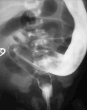 A frontal view from contrast enema in a patient initially given a diagnosis of small left colon syndrome. A long filling defect is seen in the rectosigmoid with gradual transition to a more dilated proximal bowel. The infant failed to improve, and rectal biopsy confirmed Hirschsprung disease.
Rare disorders that may partially simulate meconium plug syndrome include neuronal intestinal dysplasia, visceral neuropathies, and megacystis-microcolon-intestinal hypoperistalsis syndrome, also termed Berdon syndrome. However, radiographic and clinical features in these disorders usually are distinguished readily from meconium plug syndrome. A small (micro or mini) colon characterizes many of these diseases.
A more common problem is an infant with sepsis or metabolic disorder who presents with a nonobstructive ileus. In these patients, the intestinal dilatation resolves once the primary problem is treated.
The main problem in differential diagnosis, after the contrast enema has been performed, is Hirschsprung disease. The enema findings in neonatal Hirschsprung disease are not distinguishable from meconium plug syndrome. The most important point is the infant's response to supportive care and enemas. (See the image below.)
A frontal view from contrast enema in a patient initially given a diagnosis of small left colon syndrome. A long filling defect is seen in the rectosigmoid with gradual transition to a more dilated proximal bowel. The infant failed to improve, and rectal biopsy confirmed Hirschsprung disease.
Evaluate any infant with apparent meconium plug syndrome for Hirschsprung disease (rectal-suction biopsy) and other possible underlying disorders when findings persist after 1-2 enemas.
The normal infant's intestinal gas pattern often appears "gassy" by adult criteria. Typically, the width of a bowel loop does not exceed the width of one of the patient's lumbar vertebral bodies. Rectal gas often may be absent in the normal infant because the rectum is dependent and filled with meconium when the patient is supine. With the exception of the rectum, the colon and small bowel usually cannot be differentiated on plain film.
-
Supine frontal view of the abdomen in a newborn with meconium plug syndrome demonstrates multiple dilated loops of bowel but no rectal gas.
-
A frontal view from contrast enema in a patient initially given a diagnosis of small left colon syndrome. A long filling defect is seen in the rectosigmoid with gradual transition to a more dilated proximal bowel. The infant failed to improve, and rectal biopsy confirmed Hirschsprung disease.
-
A lateral view from contrast enema in a newborn demonstrates a normal-to-decreased caliber "empty" distal colon and dilated proximal bowel containing multiple plugs. The child responded clinically and radiographically to a single enema.
