Continually Updated Clinical Reference
 
 
  All Sources     eMedicine     Medscape     Drug Reference     MEDLINE
 
eMedicine - Appendicitis : Article by

Quick Find
Authors & Editors
Introduction
Differentials
Radiograph
CT SCAN
MRI
Ultrasound
Nuclear Medicine
Intervention
Multimedia
References

Related Articles
Cholecystitis, Acute

Cholelithiasis

Colon, Diverticulitis

Endometrioma/Endometriosis

Mesenteric Adenitis

Nephrolithiasis/Urolithiasis

Ovarian Torsion

Pelvic Inflammatory Disease/Tubo-ovarian Abscess


Patient Education
Esophagus, Stomach, and Intestine Center

Appendicitis Overview

Appendicitis Causes

Appendicitis Symptoms

Appendicitis Treatment

Abdominal Pain in Adults Overview

Abdominal Pain in Children Overview


Author: Lutfi Incesu, MD, Professor, Department of Radiology, Ondokuz Mayis University School of Medicine; Chief, Neuroradiology and MR Unit, Department of Radiology, Ondokuz Mayis University Hospital, Turkey

Lutfi Incesu is a member of the following medical societies: American Society of Neuroradiology and Radiological Society of North America

Coauthor(s): Caroline R Taylor, MD, Associate Professor, Department of Diagnostic Imaging, Yale University School of Medicine; Chief, Diagnostic Imaging Service, Department of Radiology, VA Connecticut Healthcare System

Editors: Bernard D Coombs, MB, ChB, PhD, Consulting Staff, Department of Specialist Rehabilitation Services, Hutt Valley District Health Board, New Zealand; Udo P Schmiedl, MD, PhD, Clinical Professor, Department of Radiology, University of Washington; Consulting Staff, Swedish Medical Center, University of Washington Medical Center, Seattle Radiologists; Robert M Krasny, MD, Consulting Staff, Department of Radiology, The Angeles Clinic and Research Institute; Eugene C Lin, MD, Consulting Staff, Department of Radiology, Virginia Mason Medical Center

Author and Editor Disclosure

Synonyms and related keywords: acute appendicitis, inflammation of the appendix, appendicolith, arrowhead sign, acute abdomen


Background

The appendix is a blind-ending tubular structure arising from the cecum. Appendicitis results from an acute inflammation of the appendix and creates the most common abdominal surgical emergency.

A diagnosis of acute appendicitis is usually made on the basis of a patient's clinical history in conjunction with physical examination and laboratory studies. Because the surgical aim is to operate earlybefore appendiceal rupture and peritonitis developpatients who present with typical findings undergo immediate surgery without radiologic evaluation. However, such imaging is advisable in patients with atypical symptoms, which can occur in infants and small children, the elderly, and young women.

For excellent patient education resources, visit eMedicine's Esophagus, Stomach, and Intestine Center. Also, see eMedicine's patient education articles, Appendicitis, Abdominal Pain in Adults, and Abdominal Pain in Children.

Pathophysiology

Obstruction of the narrow appendiceal lumen is the primary cause of appendicitis and may often be secondary to impacted fecal material or hyperplasia of the submucosal lymphoid follicles, which is associated with respiratory disease, infectious mononucleosis, and gastroenteritis. Fecaliths result from the inspissation of fecal material and inorganic salts within the appendiceal lumen, are the most common causes of obstruction, and are present in 11-52% of patients with acute appendicitis. Appendiceal calculi are rarely observed, and their presence is usually associated with perforating appendicitis. Appendiceal lumenal obstruction may also be secondary to strictures, parasitic infection, the presence of foreign bodies, Crohn disease, primary or secondary metastatic cancer, carcinoid syndrome, kinks, and adhesions.

The stages of appendicitis

  • Early stage of appendicitis: Obstruction of the appendiceal lumen leads to mucosal edema, mucosal ulceration, bacterial diapedesis, appendiceal distention due to accumulated fluid, and increasing intraluminal pressure. The visceral afferent nerve fibers are stimulated, and the patient perceives mild visceral periumbilical or epigastric pain, which usually lasts 4-6 hours.
  • Suppurative appendicitis: Increasing intraluminal pressures eventually exceed capillary perfusion pressure, which is associated with obstructed lymphatic and venous drainage and allows bacterial and inflammatory fluid invasion of the tense appendiceal wall. Transmural spread of bacteria causes acute suppurative appendicitis. When the inflamed serosa of the appendix comes in contact with the parietal peritoneum, patients typically experience the classic shift of pain from the periumbilicus to the right lower abdominal quadrant (RLQ), which is continuous and more severe than the early visceral pain.
  • Gangrenous appendicitis: Intramural venous and arterial thromboses ensue, resulting in gangrenous appendicitis.
  • Perforated appendicitis: Persisting tissue ischemia results in appendiceal infarction and perforation. Perforation can cause localized or generalized peritonitis.
  • Phlegmonous appendicitis or abscess: An inflamed or perforated appendix can be walled off by the adjacent greater omentum or small-bowel loops, resulting in phlegmonous appendicitis or focal abscess.

Rare types of appendicitis include the following:

  • Spontaneously resolving appendicitis: If the obstruction of the appendiceal lumen is relieved, acute appendicitis may resolve spontaneously.1, 2 This occurs if the cause of the symptoms is lymphoid hyperplasia or when a fecalith is expelled from the lumen.
  • Recurrent appendicitis: The incidence of recurrent appendicitis is 10%. The diagnosis is accepted as such if the patient underwent similar occurrences of RLQ pain at different times that, after appendectomy, were histopathologically proven to be the result of an inflamed appendix.
  • Chronic appendicitis: Chronic appendicitis occurs with an incidence of 1% and is defined by the following: (1) the patient has a history of RLQ pain of at least 3 weeks’ duration without an alternative diagnosis; (2) after appendectomy, the patient experiences complete relief of symptoms; (3) histopathologically, the symptoms were proven to be the result of chronic active inflammation of the appendiceal wall or fibrosis of the appendix.
Infected thrombi rarely form in the portal vein; when they do, pylephlebitis results.

The incidence of appendicitis is higher in more affluent countries that have a prevalence of low-fiber diets. Low-fiber diets change the bacterial flora, increasing stool viscosity, bowel transit time, and intraluminal pressure, which encourage the formation of fecaliths. These diets contribute to low-residue stool, which can become impacted within the appendiceal lumen.

Frequency

United States

In the United States, 250,000 cases of appendicitis are reported annually, representing 1 million patient-days of admission. The incidence of acute appendicitis has been declining steadily since the late 1940s, and the current annual incidence is 10 cases per 100,000 population. The overall lifetime risk of developing appendicitis is approximately 7%. Some familial predisposition exists.

International

Appendicitis appears to be a disease of prosperous Western nations; the incidence of appendicitis is higher in more affluent countries that have prevalent low-fiber diets.

Mortality/Morbidity

Acute appendicitis is the most common reason for emergency abdominal surgery. Appendectomy carries a complication rate of 4-15%, as well as associated costs and the discomfort of hospitalization and surgery. Therefore, the goal of the surgeon is to make an accurate diagnosis as early as possible. Delayed diagnosis and treatment account for much of the mortality and morbidity associated with appendicitis.

The overall accuracy for diagnosing acute appendicitis is approximately 80%, which corresponds to a mean false-negative appendectomy rate of 20%. Diagnostic accuracy varies by sex, with a range of 78-92% in male patients and 58-85% in female patients. 

  • False-negative and -positive rates associated with appendectomy reflect the diagnostic difficulty encountered in differentiating appendicitis from other acute abdominal conditions. Appendicitis may be extremely difficult to diagnose in women of childbearing age, because symptoms of acute gynecologic conditions, such as pelvic inflammatory disease, may manifest similarly. This problem results in false-positive appendectomy rates as high as 47% in female patients aged 10-39 years. Spiral computed tomography (CT) scanning and ultrasonography (US) may lower the rate of false–negative appendectomy diagnosis and reduce the rate of perforation by shortening in-hospital delay in treatment.
  • The mortality rate for acute appendicitis is less than 1 per 100,000 persons (0.2-0.8%). The mortality rate in children ranges from 0.1-1%; in patients older than 70 years, the rate rises above 20%.
  • Appendiceal perforation is associated with increased morbidity and mortality compared with nonperforating appendicitis. The mortality risk of acute but not gangrenous appendicitis is less than 0.1%, but the risk rises to 0.6% in gangrenous appendicitis. The rate of perforation varies from 16-40%, with a higher frequency occurring in younger age groups (40-57%) and in patients older than 50 years (55-70%), in whom misdiagnosis and delayed diagnosis are common. Complications occur in 1-5% of patients with appendicitis, and postoperative wound infections account for almost one third of the associated morbidity.

Race

No racial predilection exists for appendicitis.

Sex

The male-to-female sex ratio is 3:2 in teenagers and young adults. By the time patients reach their mid-30s, males and females have similar incidence rates.

Age

Appendicitis occurs in all age groups but is rare in infants; the incidence becomes increasingly common in childhood, reaching a peak incidence in the late teenage years and early 20s. The mean age when appendicitis occurs in the pediatric population is 6-10 years. 

  • Younger children have a higher rate of perforation, with reported rates of 50-85%.
  • Lymphoid hyperplasia is observed more often among infants and adults and is responsible for the increased incidence of appendicitis in these age groups.

Anatomy

The appendix vermiformis is a thin, wormlike, tubular organ that is located at the inferior part of the cecum. In adults, a normal vermiform appendix varies in length from 5-35 cm (average 8 cm).

The appendix has no fixed position. It originates 1.7-2.5 cm below the terminal ileum, either in a dorsomedial location (most common) from the cecal fundus, directly beside the ileal orifice, or as a funnel-shaped opening (2-3% of patients). The appendix has a retroperitoneal location in 65% of patients and may descend into the iliac fossa in 31%. The difference in appendiceal position considerably changes clinical findings.

Appendiceal congenital disorders are extremely rare but occasionally reported (eg, agenesis, duplication, triplication).

The appendix has its own mesentery, which arises from a peritoneal extension that extends from the terminal ileum to the medial aspect of the cecum and appendix. This mesenteric fold contains the appendicular artery, which is a terminal branch of the ileocolic artery, and runs adjacent to the appendicular wall. Venous drainage is via the ileocolic veins and the right colic vein into the portal vein; lymphatic drainage occurs via the ileocolic nodes along the course of the superior mesenteric artery to the celiac nodes and cisterna chyli.

The appendix is contained within the visceral peritoneum that forms the serosa, and its exterior layer comprises longitudinal muscle with a deeper, interior circular muscle layer. Beneath these layers lies the submucosal layer, which contains lymphoepithelial tissue. The mucosa consists of columnar epithelium with few glandular elements and neuroendocrine argentaffin cells.

Clinical Details

A diagnosis of appendicitis can usually be made on the basis of the patient’s clinical history and physical examination. Understanding the typical clinical manifestations of appendicitis is important and helps clinicians to make an early and accurate diagnosis before appendiceal perforation occurs.

Symptoms of appendicitis may take 4-48 hours to develop. The classic history of lack of appetite and periumbilical pain that is followed by nausea, RLQ pain, and vomiting occurs in only 50-60% of patients; indeed, any combination of these symptoms may occur.

  • The patient's pain is typically colicky and is initially located periumbilically or epigastrically. The pain subsequently shifts to the RLQ (50%), where it becomes progressively more severe. The migration of pain is the most discriminating historical feature, with a sensitivity and specificity of approximately 80%.
  • Nausea (61-92%), vomiting, anorexia (74-78%), and low-grade fever are common. When vomiting occurs, it almost always follows the onset of pain.
  • Diarrhea or constipation is observed in 18% of patients.
  • Abdominal rebound tenderness (96%), pain on percussion, rigidity, and guarding are specific physical findings of acute appendicitis.
  • RLQ pain with palpation of the left lower quadrant (Rovsing sign), RLQ pain with hyperextension of the right hip (psoas sign), and RLQ pain with internal rotation of the flexed right hip (obturator sign) are rarely present with acute appendicitis.
  • Leukocytosis (>10,000/mm3) is observed in 80% of patients with acute appendicitis. However, elevated levels can be noted with other conditions, and normal white blood cell (WBC) levels are often present with appendicitis. Neutrophilia is also observed (95%).
  • It has been suggested that high levels of C-reactive protein (>0.8 mg/dL) in association with leukocytosis and neutrophilia are the most sensitive laboratory findings for appendicitis, with a sensitivity of approximately 97-100%.3, 4 Therefore, the probability of acute appendicitis is low in the absence of these 3 laboratory findings.
  • A urine test may be performed to exclude urinary tract infection as the cause of the patient’s symptoms.
Despite the familiarity of these signs and symptoms of appendicitis, 20% of patients present atypically. Such atypical clinical presentations can occur in patients with a retrocecal appendix (see Images 1-2), with flank or right upper quadrant (RUQ) pain, or in patients with bowel malrotation or an elongated appendix within the pelvis or inguinal canal. The clinical diagnosis is also more problematic in infants and young children, ovulating women aged 20-40 years, and elderly patients.

Radiologic examination can reduce the number of misdiagnoses and unnecessary laparotomies, as well as help in the treatment of patients with appendiceal abscesses and those with postoperative complications.

Preferred Examination

Controversy exists as to whether imaging is required in patients with the classic history and physical findings of acute appendicitis. Opinion varies as to whether these modalities should be performed in all patients with suggested appendicitis or if radiology should be reserved for select patients with atypical or confusing clinical presentations.

Helical CT scanning and graded-compression Doppler US are powerful imaging methods that substantially improve diagnostic accuracy in patients with clinically equivocal appendicitis (see Images 1 and 3).5

Before the 1980s, abdominal radiographs and barium enema were the primary radiologic methods used in the diagnosis of acute appendicitis. On plain radiographic films, the presence of an appendicolith is the most specific sign, but it is rarely observed. On barium enema examination, nonfilling or incomplete filling of the appendix indicates appendiceal inflammation.

Continuous improvements in imaging technology, technique, and interpretation that have been achieved over the past 15 years have substantially increased the accuracy of imaging methods. Since 1986, US and, after the 1990s, CT scanning have gained acceptance as the primary imaging techniques for acute appendicitis by virtue of their ability to directly image the appendix, adjacent fat, and gut.

Graded-compression US of the RLQ has been shown to be a useful examination because of this technique's safety and high accuracy (approximately 90%) in the diagnosis of acute appendicitis. Advantages of US include lack of radiation exposure, noninvasiveness, short acquisition time, and the potential for diagnosis of other causes of abdominal pain, particularly in the subset of patients who are women of childbearing age. Several authors suggest that US should be the first imaging method used in pregnant women and pediatric patients because x-ray exposure is especially undesirable in these groups.

Contrast-enhanced, thin-section (0.5 mm) helical CT scanning has become the preferred imaging technique in the diagnosis of acute appendicitis and its complications, with a high diagnostic accuracy of 95-98% (see Image 3). The literature suggests that limited helical CT scanning with rectal contrast is a highly accurate, time-efficient, cost-effective way to evaluate adult patients with equivocal presentations for appendicitis. CT scanning is particularly preferred in patients in whom appendiceal perforation is suspected, because the diagnostic accuracy remains high and because CT scanning is useful for characterizing periappendiceal inflammatory masses.

Other advanced radiologic examinations, such as magnetic resonance imaging (MRI), scintigraphy, and color Doppler US, have been used in the diagnosis of acute appendicitis (see Images 1 and 3-5), with a diagnostic accuracy of approximately 91-95%. Currently, no practical role exists for MRI and scintigraphy in acute appendicitis. Advantages of MRI include better visualization of abnormal appendices and adjacent inflammatory processes, visualization of the appendix in an atypical location, delineation of pathology, operator independence, and ease of examination of patients who are obese. MRI, similar to enhanced CT scanning, can demonstrate the extent of inflammatory infiltration (see Image 6).

The decision to obtain US or CT scan studies depends on institutional preference and the available user expertise, although patient age, sex, and body habitus are important influencing factors. US and CT scanning have similar diagnostic value for detecting an alternative diagnosis in a patient in whom acute appendicitis is suspected. Balthazar et al showed that CT scanning is superior to US in the diagnosis of acute appendicitis6; however, US is rapid, safe, inexpensive, and noninvasive, and it requires no contrast material or patient preparation.

At the author's institution, graded-compression US and color and power Doppler US techniques are routinely used in the diagnosis of acute appendicitis, with a diagnostic accuracy of approximately 95-100% in all patients with suspected acute appendicitis. Helical CT scans or MRIs are complementary to US and are used for patients in whom the US findings are equivocal or suboptimal.

Limitations of Techniques

Abdominal radiographs are normal in many patients with acute appendicitis and should not be obtained routinely. An appendicolith is the most specific sign on plain radiographic films, but it is observed in only 10% of patients with appendicitis. Disadvantages of barium enema include a high incidence of nondiagnostic examinations, radiation exposure, insufficient sensitivity, and invasiveness.

A significant disadvantage of US is that it is operator dependent. Intestinal peristalsis, pulsations of the iliac artery (when it is close to the appendix), deep respiration in noncooperative patients, and difficulty maintaining the probe at the same location for a long time are disadvantages of color Doppler US in detecting increased vascularity of the appendix.

Disadvantages of CT scanning include radiation exposure, the potential for anaphylactoid reaction if intravenous (IV) contrast is used, lengthy preparation time if oral contrast is used, and patient discomfort if rectal contrast is used.

Disadvantages of MRI include high cost, use of IV contrast, the requirement that patients fully cooperate, difficulty with patients who are claustrophobic, the inability to observe an appendicolith in the lumen (an important finding in acute appendicitis), and the inability to differentiate between gas and an appendicolith in the perforation site.

Disadvantages of radionuclide scanning include long acquisition times (approximately 5 h) and the lack of availability of this modality.


Cholecystitis, Acute
Cholelithiasis
Colon, Diverticulitis
Endometrioma/Endometriosis
Mesenteric Adenitis
Nephrolithiasis/Urolithiasis
Ovarian Torsion
Pelvic Inflammatory Disease/Tubo-ovarian Abscess

Other Problems to Be Considered

Crohn disease
Appendagitis
Diverticulitis
Segmental omental infarction
Meckel diverticulitis
Perforated peptic ulcer
Ileal or cecal perforation
Tuberculous ileitis
Yersinial ileitis
Omental torsion
Familial Mediterranean fever


Findings

Abdominal radiographs are normal in many patients with acute appendicitis. Therefore, plain radiographic films rarely add to the diagnosis. In one study, plain-film findings consistent with acute appendicitis were observed in only 20% of patients with appendicitis.

The presence of a calcified appendiceal fecalith, which is the most specific plain-film sign, occurs in fewer than 10% of patients.

Other plain-film findings include convex lumbar scoliosis, obliteration of the right psoas margin, RLQ air-fluid levels, air in the appendix, and pneumoperitoneum.

Barium enema can be performed on an unprepared bowel quickly and safely by using the single-column technique. Complete filling of a normal appendix effectively excludes the diagnosis of appendicitis; however, nonfilling or incomplete filling of the appendix, coupled with pressure effect or spasm in the cecum, suggests appendicitis (see Image 7).

Degree of Confidence

The consensus in the literature is that plain radiography is insensitive and nonspecific. Plain radiographs should not be obtained routinely.

False Positives/Negatives

Appendicoliths may be found in individuals without appendicitis, and other plain-film signs of appendicitis can be observed in other abdominal diseases.

Although the diagnostic accuracy of a barium enema is reported to be 80-100%, this technique has several drawbacks, such as nonfilling of the appendix, which can be observed in 15-20% of patients without appendicitis.


Findings

CT scanning has the advantage of direct visualization of the appendix as well as the periappendiceal and other intra-abdominal structures. CT scanning can be used to evaluate an abnormal appendix and the nature, severity, and extent of the associated inflammatory process.

CT scanning techniques

In evaluating patients with suspected appendicitis, a number of techniques have been advocated, including abdominal and pelvic CT scanning without oral or IV contrast (see Image 3), focused CT scanning of the cecum after administration of rectal contrast, or use of oral or IV contrast, with a delay of 45 minutes to 1 hour following oral contrast administration to permit passage of the contrast into the colon.

Patient acceptance of retrograde instillation of barium per rectum may limit the use of this focused technique. A limited scanning volume may be more appropriate in young, particularly female, patients in consideration of reducing radiation exposure to the internal and reproductive organs.

Visualization of the appendix is optimized in CT scanning with use of bowel opacification, IV contrast, fine collimation (<5 mm), and/or thin-section reconstruction. The postacquisition reformatting capabilities of multislice technology should limit the necessity for rescanning a nonvisualized or poorly visualized appendix, although at a somewhat higher cost in the initial radiation dose.

CT scan findings in the normal appendix

The appendix can be visualized in approximately 50% of patients without appendicitis, although a higher visualization rate is expected in patients who are scanned with thin-section, multislice CT scanning technology (see Image 8).

The appendix is demonstrated as a tubular structure when it lies in the plane of the CT scan section or a ringlike structure when it is scanned in a cross-sectional plane. The appendix occasionally contains air or barium, but it may be collapsed. The wall thickness should be very thin, and the normal external diameter is usually less than 6 mm, although this may be slightly greater in patients with elongated retrocecal appendices.

CT scan findings in appendicitis

With high-resolution CT scanning, an abnormal appendix can be observed, and variable CT scan findings can be evaluated in patients with acute appendicitis.

Specific CT scan findings of appendicitis include enlargement of the appendix (>6 mm in the outer diameter), enhancement of the appendiceal wall, lack of opacification in an enlarged appendix, fat stranding in the periappendiceal region, and the presence of an appendicolith within the appendix (see Images 3, 9, and 10). The appendicolith may be observed outside the appendix lumen within an inflammatory mass or in a fluid collection (see Image 10).

A few secondary findings often accompany acute appendicitis. Focal thickening of the cecum may occur. The inflammatory process can be observed to separate the cecal lumen from the base of the appendix or an appendicolith (cecal bar). Contrast medium is observed within the cecum channeling to the point of the appendiceal obstruction (arrowhead sign) (see Image 11).7

Gangrenous appendicitis is recognized as enlargement of the appendix with associated fluid and loculated air within the lumen (see Image 12).

Distal appendicitis is diagnosed when CT scanning reveals appendicitis that involves the distal appendix, with a normal-appearing proximal appendix (see Image 9).

Detection of phlegmons, abscesses, and RLQ inflammation adjacent to the cecum is strongly suggestive ofbut not pathognomonic foracute appendicitis. Perforated appendicitis is usually accompanied by pericecal phlegmon or abscess formation. Free intra-abdominal air can be observed either under the right diaphragm or in a retroperitoneal location in patients with a perforated appendix. Associated CT scan findings include extraluminal air, marked ileocecal thickening, localized lymphadenopathy, peritonitis, and small-bowel obstruction (see Image 13).

Degree of Confidence

Conventional and helical CT scanning techniques have documented high accuracy (96-98%), sensitivity (96-100%), specificity (95-97%), positive predictive value (97-99%), and negative predictive value (88-100%).

CT scanning is readily available, operator independent, and relatively easy to perform, and it provides results that are easy to interpret. Furthermore, CT scanning helps clinicians to diagnose other intra-abdominal conditions in 50-80% of patients who do not have acute appendicitis (see Images 14-15).

CT scanning has high diagnostic accuracy in detecting complications, particularly in patients in whom appendiceal perforation is suspected. This modality can help clinicians to accurately differentiate mild inflammation from phlegmons and abscesses, as well identify the extent of the disease. CT scanning can be used to guide surgical or percutaneous abscess drainage.

False Positives/Negatives

Some CT scan findings that are suggestive of or often accompany acute appendicitis are nonspecific and can be observed with other RLQ conditions, such as Crohn disease (see Image 14), ulcerative colitis, pancreatitis (see Image 15), perforation of a duodenal ulcer, and cholecystitis. Misinterpretation of these signs as indicative of appendicitis can lead to a false-positive result.

Conversely, specific signs can be misapplied. Without optimal cecal opacification, a distended, inflamed appendix can be mistaken for a small-bowel loop. Rarely, an appendiceal lumen filled with appendicoliths can mimic an opacified lumen on CT scans.

Distal appendicitis can potentially be the cause of a false-negative interpretation.


Findings

Two studies regarding the use of MRI in evaluating acute appendicitis support different techniques.

A study by Incesu et al described significant enhancement of the inflamed appendix and surrounding fat (see Images 4 and 16) on gadolinium-enhanced, fat-suppressed, T1-weighted, spin-echo images.8 Mild enhancement was observed in the normal appendix and gut (see Image 17). Using fat-saturation techniques, contrast differences were observed between the inflamed appendix and the surrounding fat. Fat-suppressed, T2-weighted axial and coronal images also helped in the detection and evaluation of appendicitis and its complications.

In another study, Hormann et al obtained axial T1-weighted, turbo spin-echo sequences; T2-weighted, turbo spin-echo sequences in the axial and coronal planes; and fat-suppressed short inversion-time, inversion-recovery, turbo spin-echo sequences in the axial plane.9 On T2-weighted, ultraturbo spin-echo images, acute appendicitis demonstrated a markedly central hyperintensity and markedly hyperintense periappendiceal tissue, as well as a slightly hyperintense thickened wall. The authors suggested that unenhanced axial T2-weighted spin-echo imaging was the most sensitive sequence for the diagnosis of acute appendicitis.

Gadolinium-based contrast agents (gadopentetate dimeglumine [Magnevist], gadobenate dimeglumine [MultiHance], gadodiamide [Omniscan], gadoversetamide [OptiMARK], gadoteridol [ProHance]) have recently been linked to the development of nephrogenic systemic fibrosis (NSF) or nephrogenic fibrosing dermopathy (NFD). For more information, see the eMedicine topic Nephrogenic Fibrosing Dermopathy. The disease has occurred in patients with moderate to end-stage renal disease after being given a gadolinium-based contrast agent to enhance MRI or MRA scans. As of late December 2006, the FDA had received reports of 90 such cases. Worldwide, over 200 cases have been reported, according to the FDA. NSF/NFD is a debilitating and sometimes fatal disease. Characteristics include red or dark patches on the skin; burning, itching, swelling, hardening, and tightening of the skin; yellow spots on the whites of the eyes; jointstiffness with trouble moving or straightening the arms, hands, legs, or feet; pain deep in the hip bones or ribs; and muscle weakness. For more information, see the FDA Public Health Advisory or Medscape.

Degree of Confidence

Fat-suppressed, gadolinium-enhanced MRI images are sensitive (97%) and accurate (95%) in the detection of acute appendicitis8; however, this technique is not routinely used in the diagnosis of acute appendicitis. Unenhanced MRI scanning is also used for the evaluation of appendicitis with an accuracy of 100%.9 When the results of Hormann et al9 and Incesu et al's8 studies are compared, unenhanced MRI is found to be the more appropriate imaging modality.

False Positives/Negatives

False-negative results with MRI usually depend on technique-related limitations, such as inefficient fat saturation that causes the appendiceal wall enhancement to be obscured by mesenteric fat.

Inflammatory diseases of the gut, such as ileal diverticular abscess and inflamed ileal segments of Crohn disease that may mimic appendicitis, have been reported as false-positive results on enhanced MRI.


Findings

US is a widely available and inexpensive modality that has the potential for highly accurate imaging in the patient with suspected acute appendicitis.

With the introduction of graded-compression US by Puylaert in 1986,10 the role and use of this modality in acute appendicitis have increased dramatically.

Ultrasound technique

US examination should begin with a thorough evaluation of both the abdominal and pelvic organs.

In women with low pelvic tenderness in whom the appendix is not visualized on transabdominal US or in whom the diagnosis is not evident after abdominal and pelvic examinations have been performed, add an endovaginal US examination. Pelvic appendicitis is best demonstrated in women with endovaginal US; a posterolateral approach is suggested to evaluate the retrocecal area for retrocecal appendicitis.

Puylaert described the use of a high-frequency linear transducer as applying gentle but firm pressure in the RLQ to displace any intervening bowel gas and to decrease the distance between the transducer (5 MHz) and the appendix, thereby improving the image quality.10

Graded-compression US allows successful examination of a patient who may have peritoneal irritation and sensitivity. During the compression, maximal tenderness of an abdominal point can provide an important diagnostic clue, which is often useful in focusing the US examination on the correct area in a patient with suspected appendicitis.

US findings

  • Major US findings of acute appendicitis in the RLQ include the following:

    • An aperistaltic, noncompressible, blind-ended, sausage-shaped structure that arises from the base of the cecum

    • Distinct appendiceal wall layers

    • An outer diameter greater than 6 mm

    • A target appearance

    • Appendicolith(s)

    • Periappendiceal fluid collection

    • Echogenic prominent pericecal fat
  • Normal appendix: A normal appendix is infrequently observed using gray-scale US, but this structure can be visualized as a blind-ended, tubular, compressible intestinal loop that is continuous with the cecum and has a diameter of less than 6 mm, particularly in thin patients (see Image 18).
  • Suppurative appendicitis
    • The longitudinal US view demonstrates a nonperforated inflamed appendix that is characterized by an aperistaltic, noncompressible, blind-ended, tubular structure with a laminated wall that arises from the base of the cecum. When the inflammation is mild and visualization is optimal, 5 distinct appendiceal wall layers can be identified (see Image 19).

    • In the transverse view, the abnormal appendix often has a target appearance and its outer diameter is in excess of 6 mm in the RLQ. Researchers have studied the thickness of the appendix wall: walls surpassing 3 mm in thickness have been evaluated as pathologic.

    • An appendicolith, which is identified as an intraluminal echogenic focus with posterior shadowing, can be detected by US in approximately 30% of patients with acute appendicitis (see Images 20-21).
  • Gangrenous appendicitis: Generalized or focal loss of the echogenic submucosal layer of the appendical wall as well as the prominent surrounding echogenic fat is consistent with gangrenous appendicitis (see Images 22-23).

  • Perforated appendicitis: In the diagnosis of a perforated appendicitis, gray-scale US is also a valuable diagnostic tool, despite the fact that the perforated appendix may not be visualized in the RLQ. Irregularity and damage of the contour of the appendix by the presence of periappendiceal fluid and hyperechoic prominent pericecal fat are diagnostic of perforation (see Image 24). Gas bubbles occur within a fluid collection in cases of perforation or as a result of gas-forming organisms. A localized perforation of the appendiceal tip may also demonstrate gas pockets in the perforation site.

  • Periappendiceal phlegmon and abscess: A phlegmon appears as a localized fluid collection, which is walled off by the adjacent greater omentum and small-bowel loops (see Image 20). An appendiceal abscess appears as a complex hypoechoic mass adjacent to the cecum or appendix. In these patients, the inflamed appendix may not be visualized (see Image 25).

  • Alternative disease: US examination is also useful for diagnosing alternative pathology, such as a tubo-ovarian abscess, ovarian torsion, ovarian cyst, or mesenteric adenitis, especially in women of childbearing age. Establishment of an alternative diagnosis does not exclude appendicitis. However, 2 benefits occur when appendicitis can be excluded by US and an alternative diagnosis is made.
Color Doppler US findings

Color Doppler US is beneficial in the evaluation of inflammatory conditions of the intestinal tract, and according to most authors, this modality is a useful adjunct to conventional US in the assessment of acute appendicitis.

  • A normal appendix seldom shows findings of mild hyperemia on Doppler US examination. However, an inflamed appendix uniformly shows greater flow than a normal appendix, and circumferential color in the wall of the inflamed appendix as observed on color Doppler US images is a strong indicator of acute appendicitis (see Image 1). A flowing appearance with Doppler US has been reported as suggestive of a pathologic appendix, but absence of flow cannot differentiate a normal from an abnormal appendix.

  • Peripheral vascularity of the inflamed mesentery and omentum can be demonstrated; however, detection of hyperemia in the appendix wall is not possible in patients with gangrenous appendicitis because of the vascular necrosis adjacent to the appendix wall that has resulted from inflammation.

  • In a study by Patriquin et al, resistive index (RI) values were measured11; in normal appendix events, end-diastolic flows were found to be low, and color Doppler US signals were rare (RI = 0.85-1.00). In acute, uncomplicated appendix events, end-diastolic flows were high (RI = 0.40-0.77; mean 0.54), and color Doppler US signals were demonstrated in abundance.

Degree of Confidence

Numerous studies have documented that, in experienced hands, US has a sensitivity of 75-90%, a specificity of 86-100%, an accuracy of 87-96%, a positive predictive value of 91-94%, and a negative predictive value of 89-97% for the diagnosis of acute appendicitis. However, US is extremely operator dependent.

One author reported the ability to identify a normal appendix in as many as 82% of patients without appendicitis. When a normal appendix cannot be demonstrated and when no pericecal fluid or echogenic mass is observed, the examination is usually considered not diagnostic for appendicitis. In the same study in which a 6-mm cutoff was used, inflammation of the appendix resulted in diameters of 6-30 mm with a sensitivity of 100% and a specificity of 68%. This particular sign of an increased outer appendiceal wall diameter is an indicator of abnormality; therefore, it is more useful in excluding rather than confirming appendicitis. A 2001 study by Rettenbacher et al evaluated several hundred patients in whom appendicitis was suspected, as well as control subjects.12 The authors determined that a range of appendiceal diameter as measured by US varied from 2-13 mm, with a diameter of greater than 6 mm in 23% of control subjects.

US examination is especially limited in obese patients or those who are in significant pain. Another important limitation of US is in the diagnosis of appendiceal perforation; in this scenario the diagnosis depends on secondary findings, such as free pericecal fluid, echogenic pericecal fat, and gas bubbles in the perforation site (see Image 24).

Color Doppler US examination increases the accuracy in the diagnosis of acute appendicitis; however, potential limitations (see Limitations of Techniques) exist in using this modality to detect acute appendicitis.

False Positives/Negatives

Several major pitfalls are involved in the overdiagnosis of appendicitis with use of US, including misinterpretation of the terminal ileum as the appendix and misinterpretation of a normal appendix as an inflamed appendix.

The appendix may be inflamed secondarily as a result of a variety of inflammatory processes. In cases of intrinsic neoplasms, the appendix may be enlarged and noncompressible.

Some false-positive US findings have resulted from spontaneously resolving appendicitis.

Fluid-filled bowel loops and fecal and gaseous distention of the colon interfere with adequate visualization of the appendiceal area. Approximately 30% of appendicitis is missed by US as the result of a retrocecal appendix. This problem may be partially overcome by scanning in the coronal plane (with the transducer parallel to the iliac wing) to visualize posterior to the cecum.

A gas-filled appendix may cause diagnostic difficulty and may be misinterpreted as either a small-bowel loop or gas in a periappendiceal abscess.


Findings

Although several investigators have explored the potential of radioactive isotope imaging in patients with acute appendicitis, no practical role currently exists for this imaging modality in this patient population.

Indium-111 (111In)–labeled leukocytes are very sensitive and specific in the detection of appendicitis, with an overall accuracy of 91-95%. However, indium is expensive, the optimal time for imaging is long (17-24 h after injection), and the technique is not always readily available.

Several techniques exist for labeling leukocytes with technetium-99 (99mTc), an inexpensive and readily available isotope. In this technique, whole blood is withdrawn, and neutrophils and macrophages are labeled with 99mTc, which is administered intravenously. Then, nuclear images of the abdomen and pelvis are obtained serially over 4 hours. Localized uptake of tracer material in the RLQ suggests appendiceal inflammation.

One study used 99mTc albumin colloid–labeled leukocytes for scanning. The reported sensitivity for this technique is 89%. Another study used Tc-99m-hexamethylpropyleneamine oxime–labeled leukocytes to scan for acute appendicitis.13 The reported sensitivity was 81% and overall accuracy was 89%. 99mTc human immunoglobulin uptake has also been suggested as an accurate and safe method in the detection of acute appendicitis (see Image 5).14 Two studies of newer labeling techniques achieved sensitivities of 98% for the presence of appendicitis.

Degree of Confidence

Scintigraphy techniques are usually completed within 5 hours, a time period that can delay operative treatment longer than the delay occasioned by the use of US and CT scanning. Although future studies may confirm sensitivity as high as 98%, the lack of availability and long acquisition time limit the use of nuclear medicine as a highly sensitive diagnostic screen for appendicitis in emergency departments.

False Positives/Negatives

Before the availability of obtaining oblique abdominal views, false-negative results with scintigraphy were reported. Oblique imaging eliminates the confusion that may occur with anteroposterior imaging in cases in which the inflamed appendix is located directly over major abdominal or pelvic vessels.


In patients with uncomplicated appendicitis, appendectomy is the treatment of choice, although in some patients, appendicitis may resolve with simple antibiotic treatment alone (spontaneously resolving appendicitis).1, 2 Nonoperative follow-up observation is risky because a delay in surgery can often lead to appendiceal perforation; in addition, surgery is the mandatory treatment for perforative appendicitis as affected patients are at high risk for abscess formation.

Treatment management of patients with an appendiceal mass is controversial. Patients typically benefit from US-guided or CT scanning-guided percutaneous catheter drainage because these techniques can help clinicians to differentiate areas of pus from a phlegmon and guide percutaneous drainage, when appropriate. One research group reported a success rate approaching 90% for percutaneous drainage of periappendiceal abscesses under CT scanning guidance.15

Management of patients with an appendiceal mass can usually be divided into 3 treatment categories with CT scanning:

  • Patients with a phlegmon or a small abscess: After IV antibiotic therapy, an interval appendectomy can be performed 4-6 weeks later.
  • Patients with a larger well-defined abscess: After percutaneous drainage with IV antibiotics is performed, the patient can be discharged with the catheter in place. Interval appendectomy can be performed after the fistula is closed.
  • Patients with a multicompartmental abscess: These patients require early surgical drainage.

Medical/Legal Pitfalls

  • Approximately 10% of adults who develop appendicitis are not diagnosed correctly at the first physician encounter. Patients with an equivocal examination should be kept for observation and follow-up monitoring by serial abdominal examinations. Failure to diagnose appendicitis is the leading cause of successful malpractice claims.
  • Performing a complete physical examination of the patient is important, including examination of the genitals. Symptoms and signs of testicular torsion and ectopic pregnancy overlap with appendicitis, and these conditions result in serious morbidity if not diagnosed quickly. The literature is inconsistent as to whether rectal examination is helpful in making the diagnosis of appendicitis; however, failure to perform a rectal examination is also cited frequently in successful malpractice claims.

Special Concerns

  • Infants: A delay in diagnosis is more common and a perforated appendicitis is found more often in infants (as many as 50%).
  • Children: Overall, children with appendicitis are misdiagnosed in 25-30% of cases. The most common misdiagnosis is gastroenteritis, followed by upper and lower respiratory infections. The rate of initial misdiagnosis is inversely related to the age of the patient.
  • Women of childbearing age:

    • 33% of women of childbearing age who develop appendicitis are misdiagnosed. The most frequent misdiagnosis is pelvic inflammatory disease, which can be difficult to differentiate from appendicitis. The presence of anorexia and onset of pain more than 14 days after menses favor the diagnosis of appendicitis.
    • Other pathologies that may clinically mimic appendicitis, such as ovarian cyst hemorrhage, acute salpingitis with tubo-ovarian abscess, ovarian torsion, rupture of a dermoid tumor, or a ruptured ectopic pregnancy, are commonly found. In the era of preimaging, these conditions led to a marked diagnostic disparity as high as 45% in the rates of noncontributory surgery. To enhance preoperative diagnostic accuracy, it is important to consider the appropriate use of diagnostic imaging in the evaluation of appendicitis, even in peak age groups.
  • Pregnancy: The incidence of appendicitis is the same in pregnant women as in those who are not pregnant; however, the clinical presentation varies more in gravid women because the appendix migrates in a counterclockwise direction toward the right kidney during pregnancy. Diagnostic delay with appendiceal perforation increases fetal and maternal morbidity. Aggressive evaluation of the appendix is warranted in cases of pregnancy.
  • Elderly patients: Appendicitis in patients older than 60 years accounts for 10% of appendectomies. The incidence of misdiagnosis is increased in elderly persons relative to other adult age groups; a delay in diagnosis and perforated appendicitis is found in this age group at a rate as high as approximately 90%. Consider other etiologies in an elderly patient who presents with acute RLQ pain and fever, including diverticulitis of the right colon or a redundant sigmoid, inflammatory or infectious colitis, and focal colonic ischemia. Diverticulitis is unusual in the right colon, but this condition may be recognized on imaging studies as minimal asymmetric thickening of the bowel wall, with localized pericolic inflammatory changes.


Media file 1:  
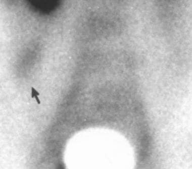
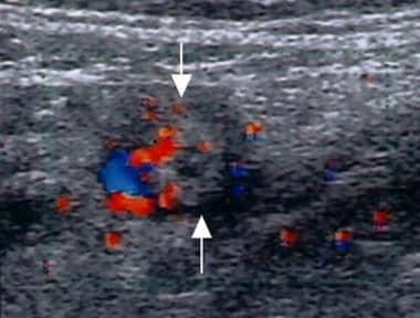
Suppurative appendicitis; transverse view, color Doppler ultrasound image. Circumferential colors are observed in the wall of the inflamed appendix (arrows), a strong indicator of acute appendicitis.
Click to see larger pictureClick to see detailView Full Size Image
Media type:  Image

Media file 2:  
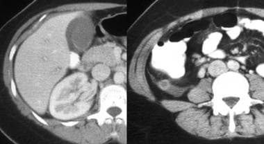
Atypical presentation of appendicitis in a young woman; computed tomography scan. The patient presented with an elevated white blood cell count and right upper quadrant pain. Left, there is pericholecystic fluid and free fluid in the right paracolic gutter, which is caused by retrocecal appendicitis. Right, the appendix, observed in axial section, has an increased diameter and an enhancing thickened wall.
Click to see larger pictureClick to see detailView Full Size Image
Media type:  CT

Media file 3:  Appendicitis in a young physician with appendicitis; computed tomography (CT) study. Intravenous (IV) contrast was administered to the patient, who had gram-negative sepsis but no abdominal pain on examination. Left, an inconclusive CT scan after administration of oral contrast but no IV contrast. Right, a repeat CT scan study following administration of IV contrast demonstrates the thickened, enhanced appendiceal wall and periappendiceal changes. The retrocecal location of the appendix may have attenuated abdominal symptoms.
Click to see larger pictureClick to see detailView Full Size Image
Media type:  CT

Media file 4:  
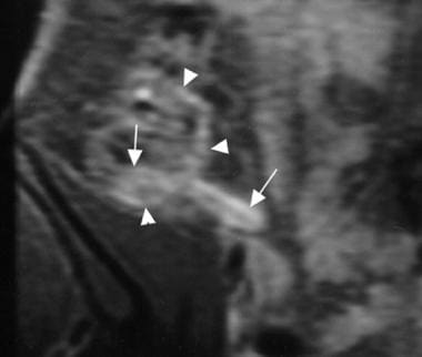
Acute suppurative appendicitis in a 15-year-old boy; contrast-enhanced, fat-suppressed, T1-weighted, spin-echo coronal magnetic resonance image. A markedly enhanced and thickened inflamed appendix (arrows) with pericecal enhancement due to the extent of inflammation is shown.
Click to see larger pictureClick to see detailView Full Size Image
Media type:  MRI

Media file 5:  Acute suppurative appendicitis in a 27-year-old woman; scintigraphy study. Pathologic accumulation of technetium-99m human immunoglobulin on the right iliac fossa is observed at 4 hours.
Click to see larger pictureClick to see detailView Full Size Image
Media type:  Image

Media file 6:  
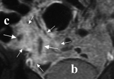
Periappendiceal abscess; contrast-enhanced, fat-suppressed, T1-weighted, spin-echo coronal magnetic resonance image. Fluid collections (long arrows) and a markedly enhanced pericecal area (short arrows) are shown. b = bladder; c = cecum.
Click to see larger pictureClick to see detailView Full Size Image
Media type:  MRI

Media file 7:  Normal appendix; barium enema radiographic examination. A complete contrast-filled appendix is observed (arrows), which effectively excludes the diagnosis of appendicitis.
Click to see larger pictureClick to see detailView Full Size Image
Media type:  X-RAY

Media file 8:  Normal appendix; computed tomography (CT) scan. A normal appendix, visualized here at the base of the cecum (arrow), is observed in 44-51% of patients. Thin-section CT scans (5-mm collimation or less) are more useful in identifying the appendix. Oral or rectal contrast should be administered. Intravenous contrast is useful in enabling enhancement and edema of the appendiceal wall to be identified.
Click to see larger pictureClick to see detailView Full Size Image
Media type:  CT

Media file 9:  Distal appendicitis; computed tomography scan. The appendiceal lumen may be normal proximally (left, arrow), but distention and inflammatory changes are noted distally (right, open arrow).
Click to see larger pictureClick to see detailView Full Size Image
Media type:  CT

Media file 10:  Retrocecal appendix; computed tomography scan. Left, the appendix occurs in a retrocecal location in 65% of patients. Right, in this young female, the appendix extends cranially as far as the posterior lobe of the liver. Appendicitis in a patient with a retrocecal appendix may present atypically, with less or poorly localized pain, discomfort on coughing or walking, or flank, rather than right lower quadrant, tenderness.
Click to see larger pictureClick to see detailView Full Size Image
Media type:  CT

Media file 11:  Arrowhead sign; computed tomography scan. The presence of this sign indicates contrast outlining the cecum and funneling into the origin of the appendix, with obstruction of the lumen preventing retrograde flow of barium into the distal appendix.
Click to see larger pictureClick to see detailView Full Size Image
Media type:  CT

Media file 12:  Acute gangrenous appendicitis with calcified appendicolith; computed tomography (CT) scan. A calcified appendicolith in the lumen of an enlarged inflamed appendix is shown.
Click to see larger pictureClick to see detailView Full Size Image
Media type:  CT

Media file 13:  Perforated appendicitis with abscess; computed tomography scan. Note the appendicolith (arrow) and air within the abscess. The terminal ileum lies anterior to the appendiceal abscess, and inflammatory change is noted in its wall, which appears thickened (open arrow).
Click to see larger pictureClick to see detailView Full Size Image
Media type:  CT

Media file 14:  Crohn disease in a young woman; computed tomography scan. The patient presented with acute right lower quadrant pain and fever. She had a normal-appearing appendix, but thickening of the wall of the terminal ileum was noted (arrow) in addition to sclerotic changes of both sacroiliac joints, that was consistent with sacroiliitis in association with Crohn disease.
Click to see larger pictureClick to see detailView Full Size Image
Media type:  CT

Media file 15:  Severe acute pancreatitis in a young man; computed tomography scans. The patient presented with peripancreatic effusions that resulted in right lower quadrant pain. Fluid had tracked down the right paracolic gutter (left, arrow, coronal section) in addition to the anterior pararenal space (right, axial view).
Click to see larger pictureClick to see detailView Full Size Image
Media type:  CT

Media file 16:  Acute suppurative appendicitis; contrast-enhanced, fat-suppressed, T1-weighted, spin-echo axial magnetic resonance image. A markedly enhanced and thickened inflamed appendix (arrows) are shown. a = iliac artery; c = cecum; p = psoas muscle; v = iliac vein.
Click to see larger pictureClick to see detailView Full Size Image
Media type:  MRI

Media file 17:  Normal appendix; contrast-enhanced, fat-suppressed, T1-weighted, spin-echo coronal magnetic resonance image. Mild enhancement in the unenlarged appendix, ileum (arrowhead), and cecum is shown.
Click to see larger pictureClick to see detailView Full Size Image
Media type:  MRI

Media file 18:  Normal appendix; longitudinal ultrasonogram. A compressible tubular appendiceal structure with an outer diameter of less than 6 mm (arrows) is shown. A = iliac artery; V = iliac vein.
Click to see larger pictureClick to see detailView Full Size Image
Media type:  Image

Media file 19:  Suppurative appendicitis in a 15-year-old boy; longitudinal ultrasonogram. An aperistaltic, noncompressible, blind-ended, fluid-filled, tubular appendiceal structure is shown, and distinct wall layers (arrows) arising from the base of the cecum are observed.
Click to see larger pictureClick to see detailView Full Size Image
Media type:  Image

Media file 20:  Phlegmonous appendicitis; oblique-axial ultrasonogram. A pericecal fluid collection, which is walled off by small-bowel loops (arrowheads) is shown, and an appendicolith with an acoustic shadow (arrow) is observed.
Click to see larger pictureClick to see detailView Full Size Image
Media type:  Image

Media file 21:  Suppurative appendicitis in a 15-year-old boy (same patient as in Image 19); longitudinal view, pathologic specimen. An inflamed appendix with appendicoliths in the lumen (arrow) is demonstrated.
Click to see larger pictureClick to see detailView Full Size Image
Media type:  Photo

Media file 22:  Gangrenous appendicitis; longitudinal ultrasonogram. A markedly distended appendix (arrows), loss of mucosa and submucosal layers, and prominent echogenic pericecal fat are shown.
Click to see larger pictureClick to see detailView Full Size Image
Media type:  Image

Media file 23:  Distended gangrenous appendix; pathologic specimen. (Same patient as in Image 22.)
Click to see larger pictureClick to see detailView Full Size Image
Media type:  Photo

Media file 24:  Perforated appendix; longitudinal ultrasonogram. A defect on the tip (large arrow, right side) of the enlarged appendix (short arrows, left side) is observed. c = cecum.
Click to see larger pictureClick to see detailView Full Size Image
Media type:  Image

Media file 25:  Periappendiceal abscess formation; oblique-axial ultrasonogram. A thick-walled, complex, hypoechoic mass adjacent to the cecum (arrows) is shown. The inflamed appendix was not visualized.
Click to see larger pictureClick to see detailView Full Size Image
Media type:  Image


  1. Migraine S, Atri M, Bret PM, et al. Spontaneously resolving acute appendicitis: clinical and sonographic documentation. Radiology. Oct 1997;205(1):55-8. [Medline].
  2. Cobben LP, de Van Otterloo AM, Puylaert JB. Spontaneously resolving appendicitis: frequency and natural history in 60 patients. Radiology. May 2000;215(2):349-52. [Medline][Full Text].
  3. Graffeo CS, Counselman FL. Appendicitis. Emerg Med Clin North Am. Nov 1996;14(4):653-71. [Medline].
  4. Gronroos JM, Gronroos P. Leucocyte count and C-reactive protein in the diagnosis of acute appendicitis. Br J Surg. Apr 1999;86(4):501-4.