Practice Essentials
The lumbar vertebrae are the 5 largest and strongest of all vertebrae in the spine. These vertebrae make up the lower back. They begin at the start of the lumbar curve (ie, the thoracolumbar junction) and extend to the sacrum. The strongest stabilizing muscles of the spine attach to the lumbar vertebrae. Fractures of lumbar vertebrae, therefore, occur in the setting of either severe trauma or pathologic weakening of the bone. Osteoporosis is the underlying cause of many lumbar fractures, especially in postmenopausal women. Osteoporotic spinal fractures are unique in that they may occur without apparent trauma. However, a thorough diagnostic workup is always required to rule out spinal malignancy. Radiography is the standard imaging study for spinal fractures. Surgical intervention is required when neurologic dysfunction and/or instability occurs as a result of a lumbar fracture. The image below reveals a wedge compression fracture. (See Pathophysiology.)
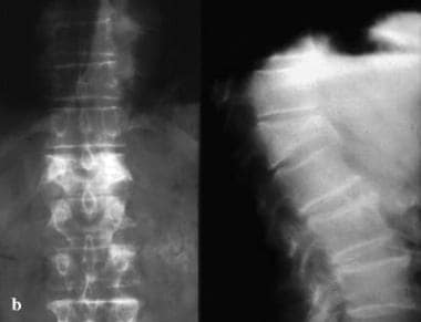 Anteroposterior and lateral radiographs of an L1 osteoporotic wedge compression fracture.
Signs and symptoms of lumbar compression fracture
Midline back pain is the hallmark symptom of lumbar compression fractures. The pain is axial, nonradiating, aching, or stabbing in quality and may be severe and disabling. The location of the pain corresponds to the fracture site, as seen on radiographs. In elderly patients with severe osteroporosis, however, there may be no pain at all as the fracture occurs spontaneously.
Diagnosis and management of lumbar compression fracture
Perform a complete blood count (CBC) with differential, prostate-specific antigen testing (in middle-aged and older men), and erythrocyte sedimentation rate determination. The urine can be sampled for markers of increased bone turnover, which occur in persons with osteoporosis.
Radiography is the standard imaging study for spine fractures. Anteroposterior and lateral views of the lumbar and thoracic spines are usually the minimum studies needed. Computed tomography (CT) scanning is an invaluable tool to evaluate the complexity of fractures seen on radiographs and to spot subtler fractures not readily seen on radiographs. Magnetic resonance imaging (MRI) is required when the patient describes lower extremity motor or sensory loss. Radicular pain is another indication for MRI. Also, when canal compromise is suspected, MRI is required. Dual-energy radiographic absorptiometry (DRA) scanning is currently the most widely used method to measure bone mineral density.
When malignancy is strongly suspected, a vertebral biopsy is indicated. These biopsies are usually carried out under CT-scan guidance. However, vertebral biopsy should not be performed when the suspected tumor is a chordoma or other aggressive primary spine tumor that spreads via direct extension.
In the past, treatment options for lumbar fractures were quite limited, with bracing and rest prescribed most often. While many patients improved with this regimen, some did not and were left with chronic, disabling pain. Suh and Lyles found that vertebral compression fractures were associated with significant performance impairments in physical, functional, and psychosocial domains in older women. [1] However, medical and surgical options are now available that can relieve the severe pain and disability from these fractures.
Traumatic injuries with neurologic compromise usually require comprehensive inpatient rehabilitation. Mobility and strength rehabilitation programs are individualized to each patient's capabilities. All therapy disciplines making up the multidisciplinary team participate in the comprehensive program. In most cases, rehabilitation begins with the patient in a thoracic-lumbar-sacral orthosis (TLSO).
Surgical intervention is required when neurologic dysfunction and/or instability occurs as a result of the lumbar fracture. The surgical procedure used for correction of a lumbar fracture depends on certain factors. These critical factors include the degree of bony canal compromise seen on axial images, the angulation on sagittal views, the level of fracture, neurologic examination findings, and the patient's premorbid health status.
Two related procedures, vertebroplasty and kyphoplasty, are available for the patient with a lumbar wedge fracture who continues to experience pain despite aggressive conservative treatment. Vertebroplasty involves injecting a form of cement polymer into the fractured vertebral body. Kyphoplasty is similar to vertebroplasty, except a balloon is used to expand the volume of the fractured segment prior to introducing the cement polymer. [2, 3, 4]
Pathophysiology
The lumbar spine provides both stability and support, allowing humans to walk upright. Proper function of the lumbar spine requires that it have a normal posture (ie, a normal lumbar curve). Any injury that changes the shape of a lumbar vertebra will alter the lumbar posture, increasing or decreasing the lumbar curve. As the body attempts to compensate for the alteration in the lumbar spine in order to maintain an upright posture, this will tend to distort the curves of the thoracic and cervical spine.
Lumbar compression fractures can be devastating injuries for 2 reasons. First, the fracture itself can cause significant pain, and this pain sometimes does not resolve. Second, the fracture can alter the mechanics of the posture. Most often, the result is an increase in thoracic kyphosis, sometimes to the point that the patient cannot stand upright. In trying to maintain their ability to walk, patients with kyphosis report secondary pain in their hips, sacroiliac joints, and spinal joints. These patients are also at risk for falls and accidents, increasing the risk of secondary fractures in the spine and elsewhere.
Fractures in the lumbar spine occur for a number of reasons. [5] In younger patients, fractures are usually due to violent trauma. Car accidents frequently cause flexion and flexion distraction injuries. Jumps or falls from heights cause burst fractures. These fractures can also result in serious neurologic injury. In older patients, lumbar compression fractures usually occur in the absence of trauma, or in the context of minor trauma, such as a fall. [6] The most common underlying reason for these fractures in geriatric patients, especially women, is osteoporosis. Other disorders that can contribute to the occurrence of compression fractures include malignancy, infections, and renal disease.
Classification of fractures
Different types of fractures can occur in the lumbar (or thoracic) spine. Classification of these fractures is based on the 3-column anatomic theory of Denis, which describes anterior, middle, and posterior spinal columns consisting of aspects of the spine and their corresponding ligaments and other soft-tissue elements. The Denis system, however, was created to classify traumatic fractures. A similar classification system does not exist for compression fractures. The main reason to use such a classification is to help determine whether a fracture is stable. Instability in the Denis system implies that damage has occurred to at least 2 of the columns of the lumbar spine.
-
Wedge fractures are the most common type of lumbar fracture and are the typical compression fracture of malignancy or osteoporosis. They occur as a result of an axially directed central compressive force combined with an eccentric compressive force. In pure flexion-compression injuries, the middle column remains intact and acts as a hinge. Although wedge fractures are usually symmetrical, 8-14% are asymmetrical and are termed lateral wedge fractures.
-
Fractures involving flexion and distraction forces are often due to lap belts in motor vehicle accidents. Commonly, the posterior columns are compromised in these injuries because the ligaments of the posterior elements are disrupted. This type of injury is quite common in young children. Most patients with flexion-distraction injuries remain neurologically intact.
-
Burst fractures result from high-energy axial loads to the spine. Multiple classification systems exist for these fractures. The severity of the deformity, the severity of canal compromise, the extent of loss of vertebral body height, and the degree of neurologic deficit affect the determination of whether these injuries are unstable.
When any of the above injuries occurs with a severe rotational force, the degree of injury and of instability increases.
Nontraumatic fractures
In osteoporosis, osteoclastic activity exceeds osteoblastic activity, resulting in a generalized decrease in bone density. The osteoporosis weakens the bone to the point where even a minor fall on the tailbone, causing an axial load or flexion, results in one or more compression fractures. The fracture is usually wedge shaped. Without correction, a wedge fracture invariably increases the degree of kyphosis.
Malignancies that result in spinal fractures are most commonly metastases rather than primary bone cancers. Primary cancers that often spread to the spine via hematologic dissemination include cancers of the prostate, kidneys, breasts, and lungs. Melanoma is a less common but more aggressive cause of spinal metastasis. The most common primary cancer of the spine is multiple myeloma, but others, including a variety of sarcomas, [7] can also manifest as a spinal fracture. Nonmalignant lesions that can cause fractures include aneurysmal bone cyst and hemangioma.
Spinal infections usually start in the lumbar intervertebral disk. From the disk, the infection spreads to bone, resulting in osteomyelitis. Severe pain is the hallmark symptom. The exception is spinal tuberculosis, or Pott disease. In this case, the disk spaces are typically spared and a compression fracture may be the initial manifestation that leads to its discovery.
A Korean study by Yoon et al found that 31.5% of elderly patients with osteoporotic vertebral compression fractures demonstrated T-score discordance between the spine and hip. The rates of major and minor discordance were 2% and 29.5%, respectively. The spinal T-score for all individuals with major discordance was lower than the femoral T-score; the same was true for 81.4% of those with minor discordance. Only age at the time of fracture was found to be associated with discordance. Mean patient age was 81.3 years. [8]
Epidemiology
Frequency
United States
The number of vertebral compression fractures occurring in the United States is estimated to be 1-1.5 million annually. While 60-75% of such fractures reportedly occur in the spine’s T12-L2 region, the L2-L5 region is the site of another 30%. [9]
Mortality/Morbidity
Mortality from a lumbar fracture is rare; however, morbidity can be significant. In elderly patients with acute osteoporotic fractures, pain and prolonged bed rest can lead to multiple secondary medical complications.
In younger persons, neurologic damage from traumatic spine injuries can result in problems such as loss of lower extremity strength and sensation and loss of bowel and bladder control.
A study by Imai et al indicated that in patients with an osteoporotic hip fracture, the coexistence of a vertebral compression fracture significantly increases mortality risk. The study involved 182 patients with osteoporotic hip fracture (average age 85 years at the time of fracture), with lumbar spine radiographs revealing vertebral compression fracture in approximately 78% of these individuals. At 1-year following hip fracture, the investigators found the mortality rate to be significantly higher in patients with a coexistent vertebral compression fracture. [10]
Sex
Osteoporosis occurs primarily in postmenopausal women. Type 1 osteoporosis occurs in women aged 51-65 years and is associated with wrist and vertebral fractures. Estrogen deficiency is the main etiologic factor. Type 2 osteoporosis (senile type) is observed in women and men older than 75 years, in a 2:1 ratio of women to men.
Age
In young and middle-aged adults, most lumbar fractures are traumatic in origin. High-velocity falls can cause burst fractures, and seat-belt injuries can cause wedge fractures. As stated above, women 51-65 years old develop type 1 osteoporosis. After age 75 years, men also begin to develop type 2 osteoporosis.
-
Anteroposterior and lateral radiographs of an L1 osteoporotic wedge compression fracture.
-
Fluoroscopic view of a kyphoplasty procedure.
Tables

What would you like to print?
- Christmas: A Time for Love and... Penile Fractures
- Hip Fractures in Patients With Dementia: To Operate or Not?
- Vertebral Fractures and Myeloma: Link Is Questionable
-
 'Decapitated' Boy Saved by Surgery Team
- Heterotopic Ossification After Modern Total Hip Arthroplasty
- Common Orthopaedic Injuries in CrossFit Athletes

 Hard Hits: Blunt Force Trauma