Piette EW, Rosenbach M. Granuloma annulare: Pathogenesis, disease associations and triggers, and therapeutic options. J Am Acad Dermatol. 2016 Sep. 75 (3):467-479. [QxMD MEDLINE Link].
Piette EW, Rosenbach M. Granuloma annulare: Clinical and histologic variants, epidemiology, and genetics. J Am Acad Dermatol. 2016 Sep. 75 (3):457-465. [QxMD MEDLINE Link].
Lapidus AH, Lee S, Khandewal T, Liu ZF, Ip KH, Lin L, et al. Subcutaneous granuloma annulare: a systematic review of a rare and underdiagnosed disease. Int J Dermatol. 2024 Aug 1. [QxMD MEDLINE Link].
Penas PF, Jones-Caballero M, Fraga J, Sánchez-Pérez J, García-Díez A. Perforating granuloma annulare. Int J Dermatol. 1997 May. 36 (5):340-8. [QxMD MEDLINE Link].
Šujica A, Bartenjev MS, Bartenjev I. Successful treatment of actinic granuloma with intralesional steroid injection: a case report. Acta Dermatovenerol Alp Pannonica Adriat. 2024 Sep. 33 (3):151-153. [QxMD MEDLINE Link]. [Full Text].
Mehta LR, Rose JW. Recurrent granuloma annulare during treatment with daclizumab. Mult Scler. 2009 Apr. 15 (4):527-8. [QxMD MEDLINE Link].
Cornejo CM, Haun P, English J 3rd, Rosenbach M. Immune checkpoint inhibitors and the development of granulomatous reactions. J Am Acad Dermatol. 2019 Nov. 81 (5):1165-1175. [QxMD MEDLINE Link].
Fontecilla NM, Kittler NW, Lopez A, Yang C, Geskin L. Programmed cell death protein-1 inhibitor-induced granuloma annulare and hypertrophic lichen planus masquerading as squamous cell carcinoma. JAAD Case Rep. 2018 Aug. 4 (7):636-639. [QxMD MEDLINE Link].
Wu J, Kwong BY, Martires KJ, Rieger KE, Chung WH, Iyer GV, et al. Granuloma annulare associated with immune checkpoint inhibitors. J Eur Acad Dermatol Venereol. 2018 Apr. 32 (4):e124-e126. [QxMD MEDLINE Link].
Kakourou T, Psychou F, Voutetakis A, Xaidara A, Stefanaki K, Dacou-Voutetakis C. Low serum insulin values in children with multiple lesions of granuloma annulare: a prospective study. J Eur Acad Dermatol Venereol. 2005 Jan. 19 (1):30-4. [QxMD MEDLINE Link].
Li A, Hogan DJ, Sanusi ID, Smoller BR. Granuloma annulare and malignant neoplasms. Am J Dermatopathol. 2003 Apr. 25 (2):113-6. [QxMD MEDLINE Link].
O'Brien JP, Regan W. Actinically degenerate elastic tissue is the likely antigenic basis of actinic granuloma of the skin and of temporal arteritis. J Am Acad Dermatol. 1999 Feb. 40 (2 Pt 1):214-22. [QxMD MEDLINE Link].
De Maeseneer M, Vande Walle H, Lenchik L, Machiels F, Desprechins B. Subcutaneous granuloma annulare: MR imaging findings. Skeletal Radiol. 1998 Apr. 27 (4):215-7. [QxMD MEDLINE Link].
Shehan JM, El-Azhary RA. Magnetic resonance imaging features of subcutaneous granuloma annulare. Pediatr Dermatol. 2005 Jul-Aug. 22 (4):377-8. [QxMD MEDLINE Link].
Blume-Peytavi U, Zouboulis CC, Jacobi H, Scholz A, Bisson S, Orfanos CE. Successful outcome of cryosurgery in patients with granuloma annulare. Br J Dermatol. 1994 Apr. 130 (4):494-7. [QxMD MEDLINE Link].
Calik J, Zawada T, Sauer N, Bove T. High Intensity Focused Ultrasound (20 MHz) and Cryotherapy as Therapeutic Options for Granuloma Annulare and Other Inflammatory Skin Conditions. Dermatol Ther (Heidelb). 2024 May. 14 (5):1189-1210. [QxMD MEDLINE Link]. [Full Text].
Hartmann Schatloff D, Retamal Altbir C, Valenzuela F. The role of excimer light in dermatology: a review. An Bras Dermatol. 2024 Nov-Dec. 99 (6):887-894. [QxMD MEDLINE Link]. [Full Text].
Sniezek PJ, DeBloom JR 2nd, Arpey CJ. Treatment of granuloma annulare with the 585 nm pulsed dye laser. Dermatol Surg. 2005 Oct. 31 (10):1370-3. [QxMD MEDLINE Link].
Sliger BN, Burk CJ, Alvarez-Connelly E. Treatment of granuloma annulare with the 595 nm pulsed dye laser in a pediatric patient. Pediatr Dermatol. 2008 Mar-Apr. 25 (2):196-7. [QxMD MEDLINE Link].
Liu A, Hexsel CL, Moy RL, Ozog DM. Granuloma annulare successfully treated using fractional photothermolysis with a 1,550-nm erbium-doped yttrium aluminum garnet fractionated laser. Dermatol Surg. 2011 May. 37 (5):712-5. [QxMD MEDLINE Link].
Bronfenbrener R, Ragi J, Milgraum S. Granuloma annulare treated with excimer laser. J Clin Aesthet Dermatol. 2012 Nov. 5 (11):43-5. [QxMD MEDLINE Link].
Passeron T, Fusade T, Vabres P, Bousquet-Rouaud R, Collet-Vilette AM, Dahan S, et al. Treatment of granuloma annulare with the 595-nm pulsed dye laser, a multicentre retrospective study with long-term follow-up. J Eur Acad Dermatol Venereol. 2013 Jun. 27 (6):785-8. [QxMD MEDLINE Link].
Harth W, Linse R. Topical tacrolimus in granuloma annulare and necrobiosis lipoidica. Br J Dermatol. 2004 Apr. 150 (4):792-4. [QxMD MEDLINE Link].
Jain S, Stephens CJ. Successful treatment of disseminated granuloma annulare with topical tacrolimus. Br J Dermatol. 2004 May. 150 (5):1042-3. [QxMD MEDLINE Link].
Rigopoulos D, Prantsidis A, Christofidou E, Ioannides D, Gregoriou S, Katsambas A. Pimecrolimus 1% cream in the treatment of disseminated granuloma annulare. Br J Dermatol. 2005 Jun. 152 (6):1364-5. [QxMD MEDLINE Link].
Lopez-Navarro N, Castillo R, Gallardo MA, Alcaide A, Matilla A, Herrera E. Successful treatment of perforating granuloma annulare with 0.1% tacrolimus ointment. J Dermatolog Treat. 2008. 19 (6):376-7. [QxMD MEDLINE Link].
Grieco T, Cantisani C, Faina P, Cantoresi F, Lacobellis F, Silvestri E, et al. Tacrolimus 0.1% and granuloma annulare: description of three cases. J Eur Acad Dermatol Venereol. 2009 Dec. 23 (12):1445-6. [QxMD MEDLINE Link].
Gomez-Moyano E, Vera-Casaño A, Martinez S, Sanz A. Periorbital granuloma annulare successfully treated with tacrolimus 0.1% ointment. Int J Dermatol. 2014 Feb. 53 (2):e156-7. [QxMD MEDLINE Link].
Kuwahara RT, Naylor MF, Skinner RB. Treatment of granuloma annulare with topical 5% imiquimod cream. Pediatr Dermatol. 2003 Jan-Feb. 20 (1):90. [QxMD MEDLINE Link].
Badavanis G, Monastirli A, Pasmatzi E, Tsambaos D. Successful treatment of granuloma annulare with imiquimod cream 5%: a report of four cases. Acta Derm Venereol. 2005. 85 (6):547-8. [QxMD MEDLINE Link].
Spindler M, Berneburg M, Drexler K, Kurz B, Kögel J, Niebel D. [Clinical variables and management of disseminated granuloma annulare - monocentric retrospective analysis of 33 cases between 2021 and 2023]. Dermatologie (Heidelb). 2024 Dec 18. [QxMD MEDLINE Link].
Errichetti E, Stinco G, Pegolo E, Patrone P. Generalized Granuloma Annulare in a Cirrhotic Patient Treated with Narrowband Ultraviolet B Therapy. Indian J Dermatol. 2016 Jan-Feb. 61 (1):127. [QxMD MEDLINE Link].
Ine K, Kabashima K, Koga C, Kobayashi M, Tokura Y, Kabashima K. Eruptive generalized granuloma annulare presenting with numerous micropapules. Int J Dermatol. 2010 Jan. 49 (1):104-5. [QxMD MEDLINE Link].
Inui S, Nishida Y, Itami S, Katayama I. Disseminated granuloma annulare responsive to narrowband ultraviolet B therapy. J Am Acad Dermatol. 2005 Sep. 53 (3):533-4. [QxMD MEDLINE Link].
Samson Yashar S, Gielczyk R, Scherschun L, Lim HW. Narrow-band ultraviolet B treatment for vitiligo, pruritus, and inflammatory dermatoses. Photodermatol Photoimmunol Photomed. 2003 Aug. 19 (4):164-8. [QxMD MEDLINE Link].
Yong A, Chong WS, Pan JY. Disseminated granuloma annulare responding to narrowband UVB phototherapy. Photodermatol Photoimmunol Photomed. 2016 Mar. 32 (2):107-9. [QxMD MEDLINE Link].
Mikami E, Yanase M, Ito M, Kanzaki A, Saeki H. Generalized granuloma annulare successfully treated with narrowband ultraviolet B and anti-hepatitis C virus therapy. J Dermatol. 2016 Aug. 43 (8):975-7. [QxMD MEDLINE Link].
Pavlovsky M, Samuelov L, Sprecher E, Matz H. NB-UVB phototherapy for generalized granuloma annulare. Dermatol Ther. 2016 May. 29 (3):152-4. [QxMD MEDLINE Link].
Kerker BJ, Huang CP, Morison WL. Photochemotherapy of generalized granuloma annulare. Arch Dermatol. 1990 Mar. 126 (3):359-61. [QxMD MEDLINE Link].
Batchelor R, Clark S. Clearance of generalized papular umbilicated granuloma annulare in a child with bath PUVA therapy. Pediatr Dermatol. 2006 Jan-Feb. 23 (1):72-4. [QxMD MEDLINE Link].
Grundmann-Kollmann M, Ochsendorf FR, Zollner TM, Tegeder I, Kaufmann R, Podda M. Cream psoralen plus ultraviolet A therapy for granuloma annulare. Br J Dermatol. 2001 May. 144 (5):996-9. [QxMD MEDLINE Link].
Browne F, Turner D, Goulden V. Psoralen and ultraviolet A in the treatment of granuloma annulare. Photodermatol Photoimmunol Photomed. 2011 Apr. 27 (2):81-4. [QxMD MEDLINE Link].
Looney M, Smith KM. Isotretinoin in the treatment of granuloma annulare. Ann Pharmacother. 2004 Mar. 38 (3):494-7. [QxMD MEDLINE Link].
Schleicher SM, Milstein HJ. Resolution of disseminated granuloma annulare following isotretinoin therapy. Cutis. 1985 Aug. 36 (2):147-8. [QxMD MEDLINE Link].
Schleicher SM, Milstein HJ, Lim SJ, Stanton CD. Resolution of disseminated granuloma annulare with isotretinoin. Int J Dermatol. 1992 May. 31 (5):371-2. [QxMD MEDLINE Link].
Tang WY, Chong LY, Lo KK. Resolution of generalized granuloma annulare with isotretinoin therapy. Int J Dermatol. 1996 Jun. 35 (6):455-6. [QxMD MEDLINE Link].
Sahin MT, Türel-Ermertcan A, Oztürkcan S, Türkdogan P. Generalized granuloma annulare in a patient with type II diabetes mellitus: successful treatment with isotretinoin. J Eur Acad Dermatol Venereol. 2006 Jan. 20 (1):111-4. [QxMD MEDLINE Link].
Pasmatzi E, Georgiou S, Monastirli A, Tsambaos D. Temporary remission of disseminated granuloma annulare under oral isotretinoin therapy. Int J Dermatol. 2005 Feb. 44 (2):169-71. [QxMD MEDLINE Link].
Adams DC, Hogan DJ. Improvement of chronic generalized granuloma annulare with isotretinoin. Arch Dermatol. 2002 Nov. 138 (11):1518-9. [QxMD MEDLINE Link].
iPLEDGE Risk Evaluation and Mitigation Strategy (REMS). US Food and Drug Administration. Available at https://www.fda.gov/drugs/postmarket-drug-safety-information-patients-and-providers/ipledge-risk-evaluation-and-mitigation-strategy-rems. December 1, 2023; Accessed: December 20, 2024.
Grewal SK, Rubin C, Rosenbach M. Antimalarial therapy for granuloma annulare: Results of a retrospective analysis. J Am Acad Dermatol. 2017 Apr. 76 (4):765-767. [QxMD MEDLINE Link].
Cannistraci C, Lesnoni La Parola I, Falchi M, Picardo M. Treatment of generalized granuloma annulare with hydroxychloroquine. Dermatology. 2005. 211 (2):167-8. [QxMD MEDLINE Link].
Simon M Jr, von den Driesch P. Antimalarials for control of disseminated granuloma annulare in children. J Am Acad Dermatol. 1994 Dec. 31 (6):1064-5. [QxMD MEDLINE Link].
Piaserico S, Zattra E, Linder D, Peserico A. Generalized granuloma annulare treated with methylaminolevulinate photodynamic therapy. Dermatology. 2009. 218 (3):282-4. [QxMD MEDLINE Link].
Weisenseel P, Kuznetsov AV, Molin S, Ruzicka T, Berking C, Prinz JC. Photodynamic therapy for granuloma annulare: more than a shot in the dark. Dermatology. 2008. 217 (4):329-32. [QxMD MEDLINE Link].
Calzavara-Pinton PG, Rossi MT, Sala R, Italian Group For Photodynamic Therapy. A retrospective analysis of real-life practice of off-label photodynamic therapy using methyl aminolevulinate (MAL-PDT) in 20 Italian dermatology departments. Part 2: oncologic and infectious indications. Photochem Photobiol Sci. 2013 Jan. 12 (1):158-65. [QxMD MEDLINE Link].
Marcus DV, Mahmoud BH, Hamzavi IH. Granuloma annulare treated with rifampin, ofloxacin, and minocycline combination therapy. Arch Dermatol. 2009 Jul. 145 (7):787-9. [QxMD MEDLINE Link].
Garg S, Baveja S. Monthly rifampicin, ofloxacin, and minocycline therapy for generalized and localized granuloma annulare. Indian J Dermatol Venereol Leprol. 2015 Jan-Feb. 81 (1):35-9. [QxMD MEDLINE Link].
Steiner A, Pehamberger H, Wolff K. Sulfone treatment of granuloma annulare. J Am Acad Dermatol. 1985 Dec. 13 (6):1004-8. [QxMD MEDLINE Link].
Czarnecki DB, Gin D. The response of generalized granuloma annulare to dapsone. Acta Derm Venereol. 1986. 66 (1):82-4. [QxMD MEDLINE Link].
Wolf F, Grezard P, Berard F, Clavel G, Perrot H. Generalized granuloma annulare and hepatitis B vaccination. Eur J Dermatol. 1998 Sep. 8 (6):435-6. [QxMD MEDLINE Link].
Saied N, Schwartz RA, Estes SA. Treatment of generalized granuloma annulare with dapsone. Arch Dermatol. 1980 Dec. 116 (12):1345-6. [QxMD MEDLINE Link].
Visconti MJ, Ashack KA, Ashack RJ. Granuloma annulare: strengthening potential associations and pentoxifylline as a therapeutic option. J Dermatolog Treat. 2021 Jun. 32 (4):381-382. [QxMD MEDLINE Link].
Wong GN, Wee E, Tam M, Kelly R. Pentoxifylline as a treatment for granuloma annulare. Australas J Dermatol. 2019 Nov. 60 (4):328-331. [QxMD MEDLINE Link].
Weber HO, Borelli C, Röcken M, Schaller M. Treatment of disseminated granuloma annulare with low-dose fumaric acid. Acta Derm Venereol. 2009. 89 (3):295-8. [QxMD MEDLINE Link].
Shupack J, Siu K. Resolving granuloma annulare with etanercept. Arch Dermatol. 2006 Mar. 142 (3):394-5. [QxMD MEDLINE Link].
Kreuter A, Altmeyer P, Gambichler T. Failure of etanercept therapy in disseminated granuloma annulare. Arch Dermatol. 2006 Sep. 142 (9):1236-7; author reply 1237. [QxMD MEDLINE Link].
Antoñanzas J, Rodríguez-Garijo N, Tomás-Velázquez A, Estenaga A, Andrés-Ramos I, España Alonso A. Treatment of recalcitrant reactive granulomatous dermatitis: Granuloma annulare subtype with etanercept. Dermatol Ther. 2020 Nov. 33 (6):e14081. [QxMD MEDLINE Link].
Murdaca G, Negrini S, Magnani O, Penza E, Pellecchio M, Gulli R, et al. Update upon efficacy and safety of etanercept for the treatment of spondyloarthritis and juvenile idiopathic arthritis. Mod Rheumatol. 2018 May. 28 (3):417-431. [QxMD MEDLINE Link].
Hertl MS, Haendle I, Schuler G, Hertl M. Rapid improvement of recalcitrant disseminated granuloma annulare upon treatment with the tumour necrosis factor-alpha inhibitor, infliximab. Br J Dermatol. 2005 Mar. 152 (3):552-5. [QxMD MEDLINE Link].
Murdaca G, Colombo BM, Barabino G, Caiti M, Cagnati P, Puppo F. Anti-tumor necrosis factor-α treatment with infliximab for disseminated granuloma annulare. Am J Clin Dermatol. 2010 Dec 1. 11 (6):437-9. [QxMD MEDLINE Link].
Amy de la Breteque M, Saussine A, Rybojad M, Kramkimel N, Vignon Pennamen MD, Bagot M, et al. Infliximab in recalcitrant granuloma annulare. Int J Dermatol. 2016 Feb. 55 (2):220-2. [QxMD MEDLINE Link].
Rosmarin D, LaRaia A, Schlauder S, Gottlieb AB. Successful treatment of disseminated granuloma annulare with adalimumab. J Drugs Dermatol. 2009 Feb. 8 (2):169-71. [QxMD MEDLINE Link].
Torres T, Pinto Almeida T, Alves R, Sanches M, Selores M. Treatment of recalcitrant generalized granuloma annulare with adalimumab. J Drugs Dermatol. 2011 Dec. 10 (12):1466-8. [QxMD MEDLINE Link].
Min MS, Lebwohl M. Treatment of recalcitrant granuloma annulare (GA) with adalimumab: A single-center, observational study. J Am Acad Dermatol. 2016 Jan. 74 (1):127-33. [QxMD MEDLINE Link].
Mahmood T, Mansouri B, Menter A. Successful treatment of generalized granuloma annulare with adalimumab. Clin Exp Dermatol. 2015 Jul. 40 (5):537-9. [QxMD MEDLINE Link].
Werchau S, Enk A, Hartmann M. Generalized interstitial granuloma annulare--response to adalimumab. Int J Dermatol. 2010 Apr. 49 (4):457-60. [QxMD MEDLINE Link].
Fässler M, Schlapbach C. Granuloma annulare arising under systemic psoriasis therapy successfully treated with adalimumab. JAAD Case Rep. 2020 Sep. 6 (9):832-834. [QxMD MEDLINE Link].
Trees A, Smithem C, Bialick M, Kabigting F. Treatment of disseminated granuloma annulare with pulse therapy upadacitinib. Dermatol Online J. 2024 Oct 15. 30 (5):[QxMD MEDLINE Link].
Piontkowski AJ, Wei N, Mumtaz A, Gulati N. Ruxolitinib cream for the treatment of granuloma annulare. JAAD Case Rep. 2024 Aug. 50:62-64. [QxMD MEDLINE Link]. [Full Text].
Wang A, Singh K, Ibrahim W, King B, Damsky W. The Promise of JAK Inhibitors for Treatment of Sarcoidosis and Other Inflammatory Disorders with Macrophage Activation: A Review of the Literature. Yale J Biol Med. 2020 Mar. 93 (1):187-195. [QxMD MEDLINE Link].
Damsky W, Singh K, Galan A, King B. Treatment of necrobiosis lipoidica with combination Janus kinase inhibition and intralesional corticosteroid. JAAD Case Rep. 2020 Feb. 6 (2):133-135. [QxMD MEDLINE Link].
Damsky W, Thakral D, McGeary MK, Leventhal J, Galan A, King B. Janus kinase inhibition induces disease remission in cutaneous sarcoidosis and granuloma annulare. J Am Acad Dermatol. 2020 Mar. 82 (3):612-621. [QxMD MEDLINE Link].
Blum S, Altman D. Treatment of generalized granuloma annulare with apremilast: A report of 2 cases. JAAD Case Rep. 2019 Nov. 5 (11):976-978. [QxMD MEDLINE Link].
Bishnoi A, Raj D, Vinay K, Dogra S. Refractory Generalized Granuloma Annulare Treated With Oral Apremilast. JAMA Dermatol. 2019 Nov 1. 155 (11):1318-1320. [QxMD MEDLINE Link].
Abdin R, Pulikanti V, Issa NT. Treatment of Granuloma Annulare Using Tapinarof Cream 1. J Drugs Dermatol. 2024 Oct 1. 23 (10):889-893. [QxMD MEDLINE Link].
Gass JK, Todd PM, Rytina E. Generalized granuloma annulare in a photosensitive distribution resolving with scarring and milia formation. Clin Exp Dermatol. 2009 Jul. 34 (5):e53-5. [QxMD MEDLINE Link].
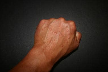 Granuloma annulare. Image from Mierlo at English Wikipedia via Wikimedia Commons. Public domain.