Practice Essentials
The epididymis is a coiled, tubular structure located along the posterior aspect of the testis. It allows for storage, maturation, and transport of sperm, connecting the efferent ducts of the testis to the vas deferens. Acute epididymitis consists of pain, swelling, and inflammation of the epididymis lasting less than 6 weeks. [1] If the testis is involved, the condition is called epididymo-orchitis. [2] Acute infective epididymitis is the most common cause of scrotal pain in adults. Active inflammation of the epididymis is seen in the ultrasound image below.
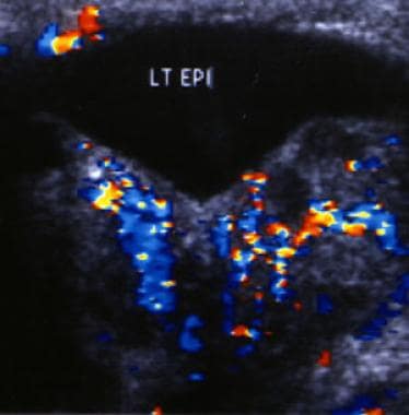 Color Doppler sonogram of the left epididymis in a patient with acute epididymitis. The image demonstrates increased blood flow in the epididymis resulting from the active inflammation.
Most cases of epididymitis occur in males aged 20 to 39 years, and most cases are associated with a sexually transmitted disease. In sexually active men older than 35 years, the most common causes are Chlamydia trachomatis and Neisseria gonorrhoeae. [1, 2] After 39 years of age, the most common causes are Escherichia coli and other coliform bacteria. In the United States, more than 600,000 men are affected annually. Reflux of urine into the ejaculatory ducts has been reported to be the most common cause of epididymitis in children younger than 14 years. [3, 1, 4]
Acute epididymitis is treated with antibiotic therapy, analgesics for pain control, and supportive care, which includes scrotal elevation and support, application of an ice pack, and, in some cases, spermatic cord block.
Diagnosis
The Centers for Disease Control and Prevention (CDC) recommends using the following tests and findings to help diagnose acute epididymitis [2] :
-
Gram or methylene blue or gentian violet stain of urethral secretions demonstrating ≥2 white blood cells (WBCs) per oil immersion field
-
Positive leukocyte esterase testing on first-void urine
-
Microscopic examination of sediment from a spun first-void urine demonstrating ≥10 WBCs per high-power field
-
Chlamydia trachomatis and Neisseria gonorrhoeae testing, preferably on urine, by nucleic acid amplification testing (NAAT)
-
Urine bacterial cultures
In a study of 237 patients with acute epididymitis, a causative pathogen was identified in 132 antibiotic-naive patients and in 44 pretreated patients. The primary pathogen was Escherichia coli. Sexually transmitted infections were present in 34 cases (25 patients with C trachomatis). [10]
Tumors, segmental testicular infarction, testicular vasculitis, pancreatitis, brucellosis, spermatic vein thrombosis, acute aortic syndrome, and appendicitis have all been identified as rare underlying causes of acute scrotal pain. [11]
Differentiation of epididymitis from testicular torsion
Patients with testicular or scrotal pain require immediate evaluation to identify and quickly treat testicular torsion, which is a true scrotal emergency. Although most cases of torsion occur in patients aged 12-18 years, testicular torsion should be considered in any patient aged 12-30 years who presents with scrotal pain. Immediate urologic consultation should be obtained if testicular torsion cannot be clearly differentiated from epididymitis or other scrotal pathology. [12, 13]
The TWIST (Testicular Workup for Ischemia and Suspected Torsion) scoring system uses clinical findings to determine the risk of testicular torsion. TWIST consists of the following parameters:
-
Testis swelling (2 points)
-
Hard testis (2)
-
Absent cremasteric reflex (1)
-
Nausea/vomiting (1)
-
High-riding testis (1)
Based on TWIST scores, patients are stratified into one of three risk groups. :
-
Low risk (0-2 points)
-
Intermediate risk (3-4 points)
-
High risk (5-7 points)
Results may be used to guide management: Prompt surgery for patients at high risk and ultrasound evaluation for intermediate-risk patients; low-risk patients do not require ultrasound to rule out torsion.
However, TWIST is necessarily imperfect: A meta-analysis concluded that in the low-risk group, for every 100 presentations of acute scrotum, 1.6 cases of testicular torsion would be missed, and in the high-risk group, surgical exploration would be negative for testicular torsion in 2.5 of 100 presentations. Other scoring systems for TWIST have been proposed (eg, risk groups of 0, 1-5, and 6-7 points), but even patients with a score of 0 may rarely have testicular torsion, and the meta-analysis concluded that the system above provides the highest reliability. [14]
Misdiagnosis of testicular torsion as epididymitis often results when reliance on imperfect diagnostic tests overrides clinical judgment. Surgical exploration to definitively exclude torsion is not a high-risk procedure. Insist on rapid, in-person consultation by the urologist in suspect cases.
Ultrasonography
Ultrasonography is noninvasive and can help to differentiate epididymitis from testicular torsion. [15, 16] (The ultrasonogram below reveals the presence of epididymitis.) One topic of study is the ability of emergency physicians to use bedside ultrasonography to accurately diagnose acute scrotal pain.
Color Doppler sonogram of the left epididymis in a patient with acute epididymitis. The image demonstrates increased blood flow in the epididymis resulting from the active inflammation.
In a retrospective chart review of 36 patients with a chief complaint of acute scrotal pain who were evaluated by an emergency physician (EP) with the aid of bedside ultrasonography, ultrasonographic findings were consistent with the results of confirmatory studies (radiology or surgery) in 35 of 36 patients. Bedside ultrasonography had a sensitivity of 95% and a specificity of 94%. [17]
In a study of 134 adult patients with acute epididymitis who underwent scrotal ultrasonography and palpation on first presentation, epididymitis was predominantly located in 24 cases (17.9%) in the head, 52 cases (38.8%) in the tail, and 58 cases (43.3%) in both. Common ultrasound features included hydrocele, epididymal enlargement, hyperperfusion, and testicular involvement. Under conservative treatment, ultrasound parameters normalized without evidence of testicular atrophy, even in patients with epididymal abscess or concomitant orchitis. [18]
However, although the use of bedside ultrasonography to accurately diagnose acute scrotal pain is promising, the skill level of EPs in using ultrasonography varies, and larger randomized, prospective, blinded studies are needed to further evaluate the accuracy of these study results.
Boettcher et al. performed a retrospective study to differentiate torsion of the appendix testis (AT) from epididymitis and found that the best predictors for epididymitis were dysuria, a painful epididymis on palpation, and altered epididymal echogenicity and increased peritesticular perfusion noted on ultrasound studies. For torsion of the AT, the best predictor was a positive blue dot sign (a tender nodule with blue discoloration on the upper pole of the testis). [19]
CRP and ESR
A study by Asgari et al suggests that C-reactive protein (CRP) levels and erythrocyte sedimentation rate (ESR) may be useful in differentiating epididymitis from testicular torsion. In this prospective study, investigators evaluated 120 patients with the diagnosis of acute scrotum; serum CRP and ESR were drawn at the time of admission. Of 46 patients with diagnosed epididymitis, 44 (95.6%) had elevation in CRP levels, and among 23 patients with torsion, 1 (4%) had an elevated CRP level. Of the 51 additional patients with other noninflammatory causes of acute scrotum, none had significant elevation in CRP levels. In addition, ESR was highest in the epididymitis group. Study authors proposed cutoff values for distinguishing epididymitis from noninflammatory causes of acute scrotum of 24 mg/L for CRP and 15.5 mm/hr for ESR. [20]
Medical Care
Emergency department care
Acute epididymitis is treated with antibiotic therapy, analgesics for pain control, and supportive care, which includes scrotal elevation and support, application of an ice pack, and, in some cases, spermatic cord block. [5, 21]
In general, antibiotics should be used in all cases of epididymitis, regardless of a negative urinalysis or the urethral Gram stain result. Nonsteroidal anti-inflammatory drugs or narcotic analgesics are also generally prescribed for patients with epididymitis. [1, 3]
Prepubertal patients require empirical coverage for coliform bacteria (enteric gram-negative bacilli or Pseudomonas). These patients may be treated with trimethoprim-sulfamethoxazole. However, in a study of epididymitis in 140 boys aged 2 months to 17 years (median age, 11 yr), only 4 of 97 patients had a positive urine culture. Given this low rate of a bacterial cause, the study authors recommend a selective approach to antibiotic therapy in pediatric epididymitis. They suggest treating all young infants, regardless of urinalysis results, and older boys who have a positive urinalysis or culture. They also recommend presumptive treatment of sexually active adolescents with epididymitis for sexually transmitted infection (STI). This study excluded boys with recent urologic surgery and known lower urinary tract anomalies. [22]
Similarly, a British study found that in prepubertal boys, a viral infection may be the most likely explanation for epididymitis. The authors note that management should be supportive and antibiotics reserved for patients with pyuria or positive cultures. [23]
In prepubertal boys, a viral infection may be the most likely explanation, according to a British study. The authors note that management should be supportive and antibiotics reserved for patients with pyuria or positive cultures. Urodynamic studies and renal tract ultrasonography are suggested for those with recurrent epididymitis. [23]
In prepubertal boys, a viral infection may be the most likely explanation, according to a British study. The authors note that management should be supportive and antibiotics reserved for patients with pyuria or positive cultures. Urodynamic studies and renal tract ultrasonography are suggested for those with recurrent epididymitis. [23]
For sexually active males, the Centers for Disease Control and Prevention (CDC) recommends presumptive therapy for STI before all laboratory test results are available, to prevent complications and transmission of STIs. [2] For acute epididymitis that is most likely caused by chlamydia or gonorrhea, the CDC recommends ceftriaxone 500 mg IM in a single dose (1 g ceftriaxone for persons ≥150 kg), plus doxycycline 100 mg orally 2 times a day for 10 days. For acute epididymitis that is most likely caused by chlamydia, gonorrhea, or enteric organisms (in men who practice insertive anal sex), the CDC recommends ceftriaxone 500 mg IM in a single dose (1 g ceftriaxone for persons ≥150 kg), plus levofloxacin 500 mg orally once daily for 10 days.
In men older than 35 years, epididymitis is usually caused by enteric bacteria (typically, Escherichia coli) introduced into the ejaculatory ducts by reflux of urine secondary to bladder outlet obstruction. [3] Enteric bacteria are also the usual pathogen in epididymitis following prostate biopsy, vasectomy, and other urinary tract instrumentation procedures. If the infection is most likely caused by enteric organisms only and if gonorrhea has been ruled out by Gram stain, methylene blue, or gentian violet stain, the CDC recommends considering levofloxacin monotherapy. [2]
Inpatient care
Most cases of epididymitis can be managed on an outpatient basis. However, the patient should be admitted for parenteral therapy if any of the following are present:
-
Intractable pain
-
Nausea or vomiting that interferes with oral therapy
-
Clinical evidence of an abscess (or inability to rule out an abscess)
-
Signs of toxicity or possible sepsis
-
Failure to improve during initial 72 hours of outpatient management
-
Immunocompromised with significant signs or symptoms
The CDC advises that the following risk factors for more severe disease may be indications for hospitalization [2] :
-
Advanced age
-
Diabetes
-
Fever
-
Elevated C-reactive protein level
Outpatient care
Treatment should be initiated as discussed above.
The patient must be evaluated by a urologist within 3-7 days of presentation. This follow-up is mandatory, as a testicular tumor occasionally is the true cause of symptoms.
The patient must receive detailed instructions for treatment, including reasons for immediate return.
Patients with epididymitis secondary to a potential sexually transmitted disease along with all of their sexual contacts need referrals for screening and diagnosis of all comorbid sexually transmitted diseases, including human immunodeficiency virus (HIV) infection.
For pediatric patients with no prior urologic history and in the absence of bacteriuria, one retrospective study suggests that investigation for underlying urinary tract pathology should be carried out if a second episode occurs. [24] Urodynamic studies and renal tract ultrasonography are suggested for those with recurrent epididymitis. [23]
-
Color Doppler sonogram of the left epididymis in a patient with acute epididymitis. The image demonstrates increased blood flow in the epididymis resulting from the active inflammation.
