Practice Essentials
Carpal tunnel syndrome (CTS) is a collection of characteristic symptoms and signs that occurs following compression of the median nerve within the carpal tunnel. Usual symptoms include numbness, paresthesias, and pain in the median nerve distribution. These symptoms may or may not be accompanied by objective changes in sensation and strength of median-innervated structures in the hand. [1, 2] See image below.
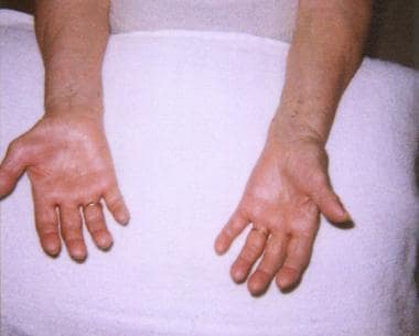 The hands of an 80-year-old woman with a several-year history of numbness and weakness are shown in this photo. Note severe thenar muscle (abductor pollicis brevis, opponens pollicis) wasting of the right hand, with preservation of hypothenar eminence.
Signs and symptoms of carpal tunnel syndrome
Among the most common complaints, patients will reveal that their hands fall asleep or that things slip from their fingers without their noticing (loss of grip, dropping things); numbness and tingling also are commonly described. Such complaints should be localized to the palmar aspect of the first to the fourth fingers and the distal palm (ie, the sensory distribution of the median nerve at the wrist).
The sensory symptoms above commonly are accompanied by an aching sensation over the ventral aspect of the wrist. This pain can radiate distally to the palm and fingers or, more commonly, extend proximally along the ventral forearm.
Not infrequently, patients report symptoms in the whole hand. Many patients with CTS also complain of a tight or swollen feeling in the hands and/or temperature changes (eg, hands being cold/hot all the time).
Pathophysiology
Until the advent of electrophysiologic testing in the 1940s, carpal tunnel syndrome (CTS) commonly was thought to be the result of compression of the brachial plexus by cervical ribs and other structures in the anterior neck region (so-called thoracic outlet syndrome). It is now known that the median nerve is damaged within the rigid confines of the carpal tunnel, initially undergoing demyelination followed by axonal degeneration. Sensory fibers often are affected first, followed by motor fibers. Autonomic nerve fibers carried in the median nerve also may be affected.
The cause of the damage is subject to some debate; however, it seems likely that abnormally high carpal tunnel pressures exist in patients with CTS. This pressure causes obstruction of venous outflow, back pressure, edema formation, and ultimately, ischemia in the nerve.
The risk of development of CTS appears to be associated, at least in part, with a number of different epidemiologic factors, including genetic, medical, social, vocational, avocational, and demographic. [20] A complex interaction probably exists between some or all these factors, eventually leading to the development of CTS. Definite causative factors, however, are far from clear.
Epidemiology
Frequency
United States
The incidence of carpal tunnel syndrome is 1-3 cases per 1000 subjects per year; prevalence is approximately 50 cases per 1000 subjects in the general population. Incidence may rise as high as 150 cases per 1000 subjects per year, with prevalence rates greater than 500 cases per 1000 subjects in certain high-risk groups.
International
A paucity of population-based studies of carpal tunnel syndrome (CTS) exists; however, the incidence and prevalence in developed countries seems similar to the United States (eg, incidence in the Netherlands is approximately 2.5 cases per 1000 subjects per year; prevalence in the United Kingdom is 70-160 cases per 1000 subjects). [21, 22, 23] CTS is almost unheard of in some developing countries (eg, among nonwhite South Africans).
Mortality/Morbidity
Carpal tunnel syndrome is not fatal, but it can lead to complete, irreversible median nerve damage, with consequent severe loss of hand function, if left untreated.
Race
Whites are probably at highest risk of developing carpal tunnel syndrome (CTS). The syndrome appears to be very rare in some racial groups (eg, nonwhite South Africans). [23] In North America, white US Navy personnel have CTS at a rate 2-3 times that of black personnel. [24]
Sex
The female-to-male ratio for carpal tunnel syndrome is 3-10:1.
Age
The peak age range for development of carpal tunnel syndrome (CTS) is 45-60 years. Only 10% of patients with CTS are younger than 31 years.
-
The hands of an 80-year-old woman with a several-year history of numbness and weakness are shown in this photo. Note severe thenar muscle (abductor pollicis brevis, opponens pollicis) wasting of the right hand, with preservation of hypothenar eminence.
-
Sensory nerve conduction studies from the left hand of a patient with a several-year history of numbness and weakness (responses from the median nerve in the right hand were completely absent). Note marked slowing of the conduction velocity (CV) to 29.8 and 25.5 m/s for digits 3 and 1, respectively (normal >50 m/s). The amplitude for both also is reduced markedly (normal >10). These findings are consistent with carpal tunnel syndrome.
-
Motor nerve conduction studies from the left hand of a patient with a several-year history of numbness and weakness (responses from the median nerve in the right hand were completely absent). Note that the conduction velocity (CV) across the carpal tunnel segment slows severely to 18.3 m/s (normal >50 m/s) and that the distal motor latency is prolonged at 6.3 ms (normal < 4.2 ms). Amplitudes are low for the wrist and elbow stimulus sites at 4.7 mV (normal >5 mV), but amplitudes are 31% higher distal to the carpal tunnel (at the palm). This discrepancy may represent conduction block (neurapraxia) at the level of the carpal tunnel or coactivation of the ulnar branch to adductor pollicis. Needle electromyography is required to determine whether axonal loss is present.
