Background
Hyponatremia is defined as a serum sodium (Na) concentration of less than 135 mEq/L. Plasma Na plays a significant role in plasma osmolality and tonicity (serum osmolarity = 2Na + Glu/18 + BUN/2.8). Changes in plasma osmolality are responsible for the signs and symptoms of hyponatremia and also the complications that happen during treatment in the presence of high-risk factors. Whereas hypernatremia always denotes hypertonicity, hyponatremia can be associated with low, normal, or high tonicity. Hyponatremia is the most common electrolyte disorder encountered in hospitalized patients. [1]
Hyponatremia can lead to hyponatremic encephalopathy, particularly in vulnerable patient populations. Low serum sodium predicts mortality and morbidity in all hospitalized patients irrespective of the severity of hyponatremia. Hospitalized patients, post-operative patients, children, and women in the reproductive age group are more vulnerable to the neurologic effects of hyponatremia.
The image below lists drugs that impair water excretion.
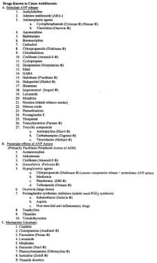 Pediatric Hyponatremia. Drugs that impair water excretion.
Clinical presentation of hyponatremia happens as a result of a rapid fall in serum Na and also the absolute level of serum Na. Fifty percent of presenting children develop symptoms when serum Na levels fall below 125 mEq/L, a relatively high level when compared with adults. Although morbidity widely varies, serious complications can arise from hyponatremia and can also happen during treatment. Understanding the pathophysiology and treatment options for hyponatremia is important because significant morbidity and mortality are possible.
Patient education
Advise parents not to replace diarrheal fluid loss with hypotonic fluids such as tea or soda.
Pathophysiology
Hyponatremia can develop because of (1) excessive free water, a common cause in hospitalized patients receiving hypotonic solutions; (2) excessive renal or extrarenal losses of Na or renal retention of free water; and, rarely, (3) deficient intake of Na.
Under normal circumstances, the human body is able to maintain serum Na in the normal range (135-145 mEq/L) despite wide fluctuations in fluid intake. The body's defense against developing hyponatremia is the kidney's ability to generate dilute urine and excrete free water in response to changes in serum osmolarity and intravascular volume status.
Hospital-acquired hyponatremia is the most common cause of hyponatremia in children. Some studies have outlined the association of hyponatremia and the hypotonic fluid typically used in the pediatric population. Excessive antidiuretic hormone (ADH) is present in most hospitalized patients, either as an appropriate response to hemodynamic and/or osmotic stimuli or as an inappropriate secretion of ADH. ADH is also secreted in response to pain, nausea, and vomiting and during the use of certain medications (such as morphine) during the postoperative period. Use of hypotonic fluids in the presence of circulating ADH can cause free-water retention, resulting in hyponatremia. In certain clinical conditions, ADH secretion occurs even when serum osmolarity is low or normal, hence the term syndrome of inappropriate ADH secretion (SIADH).
Other conditions that can lead to hyponatremia include states with increased total body water such as with cirrhosis, cardiac failure, or nephrotic syndrome. Diuretic use and decreased intake of Na can also lead to hyponatremia.
Loss of Na via the gastrointestinal (GI) tract and/or urinary tract in excess of free water can result in hyponatremia. GI losses can occur in different disease states with excessive fluid loss, namely gastroenteritis, fistulas, or serous fluid drainage after surgery. Na can be lost via the kidney; use of diuretics is the most common culprit, followed by other causes, such as salt-losing nephritis, mineralocorticoid deficiency, and cerebral salt-wasting syndrome (CSWS). As stated above, hyponatremia is rarely caused by deficient Na intake.
Clinical manifestations vary from an asymptomatic state to severe neurologic dysfunction. Central nervous system (CNS) symptoms predominate in hyponatremia, although cardiovascular and musculoskeletal findings may be present. Factors that contribute to CNS symptoms are (1) the rate at which serum Na levels change, (2) the absolute serum Na level, (3) the duration of the abnormal serum Na level, (4) the presence of other CNS pathology risk factors, (5) the presence of excessive ADH levels, (6) the age of the patient, (7) the sex of the patient, and (8) the presence of associated hypoxia.
CNS effects
Hyponatremia exerts most of its clinical effects on the brain. Brain volume is regulated by equal osmolality of extracellular and intracellular fluid.
If hyponatremia is acute (ie, within 48 hours), the change in osmolality causes influx of water, resulting in cerebral edema. (Cerebral edema is responsible for symptoms such as headache, nausea, vomiting, irritability, and seizures.) If hyponatremia occurs slowly (ie, after 48 hours), the brain has an adaptive response to protect itself from edema formation. The brain’s adaptive response is mediated through different mechanisms and also modified by different factors, as discussed below.
Mechanisms implied in cerebral edema formation include the following:
-
Na-K adenosine triphosphatase (ATPase) system
-
Aquaporin channels
-
Organic osmolytes
The influx of water into the brain that occurs in hyponatremia-associated reduced osmolarity occurs primarily through glial cells and largely via the water channel aquaporin (AQP). Water is then shunted to astrocytes, which swell, largely preserving the neurons. Na is extruded at the same time using the Na-K ATPase system. Potassium ions extrusion follows Na but is slower. In addition, inorganic osmolytes and organic osmolytes (eg, glycine, taurine, creatine, and myoinositol) have been shown to efflux from cells during hypo-osmolar states in animal studies.
The brain’s adaptive response to protect itself from edema occurs over several days. Once the brain has adapted to the hypo-osmolar conditions, a correction of the hypo-osmolar extracellular space to a euvolemic or hyper-osmolar state that is too rapid leads to a rapid efflux of water from brain tissue, resulting in dehydration of brain cells. The resultant condition is called osmotic demyelination syndrome (ODS). Previously, this pathologic injury was described only in the pons (hence the term central pontine myelinolysis [CPM]). Although it predominantly affects the pons, this condition is now known to occur in other parts of the brain as well (see Complications).
Hyponatremic encephalopathy
Risk factors for hyponatremic encephalopathy include age, sex, hypoxia, and vasopressin levels.
Sex
Epidemiologic data have shown that the risk of developing permanent neurologic sequelae or death from hyponatremic encephalopathy is substantially higher in menstruating women than in men or postmenopausal women. [2] The relative risk of death or permanent neurologic damage due to hyponatremic encephalopathy is about 30 times greater for women than for men and about 25 times greater for menstruating women than for postmenopausal women.
Although estrogen hormones have been implicated as the cause of this high incidence of hyponatremic encephalopathy, cellular-level mechanisms have now been elucidated. Estrogen has a core steroidal structure similar to cardiac glycosides known to inhibit the Na-K ATPase system, impairing adaptive responses. In addition, estrogen also appears to regulate water movement and neurotransmission by affecting AQP4 expression.
Age
Children are at increased risk of developing complications because of hyponatremia. The smaller the child, the higher the risk. Although many other factors may contribute to this increased risk, brain-to–cranial vault ratio plays an important role.
The brain reaches adult size by age 6 years, whereas the skull does not reach adult size until age 16 years. As a consequence, children can develop symptomatic hyponatremia with relatively higher Na concentrations than those observed in adults.
Good outcomes are reported in young babies with open fontanelles; increased vault compliance supports this hypothesis.
Hypoxia
Hypoxia is a major risk factor for hyponatremic encephalopathy. Patients with symptomatic hyponatremia can develop hypoxia by two different mechanisms: noncardiogenic pulmonary edema (Ayus-Arieff syndrome) and hypercapnic respiratory failure. Hypercapnic respiratory failure is due to central respiratory depression and is often the first sign of impending transtentorial herniation. Noncardiogenic pulmonary edema, on the other hand, is a complex disorder with increased vascular permeability and increased catecholamine release that often occurs secondary to elevated intracranial pressure.
Hypoxia worsens clinical outcomes in hyponatremic encephalopathy by impairing the brain’s adaptive response through the active transport of Na, which is an energy-dependent process that requires oxygen. In addition, it affects astrocyte volume regulation, which is also energy dependent. Under ordinary circumstances, hypoxia increases cerebral blood flow to increase the delivery of oxygen; [3] the increase in cerebral blood flow can lead to an increase in cerebral blood volume, which also contributes to an increase in intracranial pressure.
Vasopressin
Hyponatremia, except in cases of pure water intoxication, virtually always occurs in the presence of increased plasma levels of vasopressin. [4]
Vasopressin leads to decreased cerebral oxygen use in the female rat brain but not in male rats. Vasopressin decreases cerebral blood flow by vasoconstriction, resulting in decreased oxygen delivery that, in turn, impairs brain adaptation. Vasopressin also facilitates the direct movement of water into brain cells independent of hyponatremia. In addition, it also decreases the synthesis of adenosine triphosphate (ATP) and phosphocreatine, lowers intracellular pH and intracellular buffering, and decreases Ca2+, which affects energy-dependent processes involved in brain adaptation.
In a retrospective study of neonates and infants under age 3 months receiving vasopressin in a neonatal intensive care unit (NICU), Patel et al found that 54.3% of the patients developed hyponatremia (defined in the study as a serum Na level of 130 mEq/L or less). Among those with hyponatremia, 29.8% had a severe form of the condition. The odds of developing hyponatremia were lower in patients with a higher baseline Na level, and greater in those with a longer duration of vasopressin treatment. [5]
Cardiovascular response to hyponatremia
Hyponatremia is also often classified by body water volume status: hyponatremia in conjunction with hypervolemia, euvolemia, or hypovolemia. The distribution of water and solute in the intracellular and extracellular spaces determines the intravascular volume. Fluid shifts from the extracellular space to the intracellular space with a subsequent decrease in arterial blood volume. The reduction in intravascular volume may result in hypotension. Because of this fluid shift, hyponatremia causes hemodynamic disturbance more pronounced than that expected for the degree of dehydration.
Etiology
Hypervolemic hyponatremia (excess free-water retention)
The following are causes of hypervolemic hyponatremia:
-
Congestive heart failure
-
Cirrhosis
-
Nephrotic syndrome
-
Acute or chronic renal failure
Hypovolemic hyponatremia due to renal loss of sodium in excess of free water
The following are causes of hypovolemic hyponatremia resulting from renal sodium loss in excess of free water:
-
Diuretic excess
-
Osmotic diuresis
-
Salt-wasting diuresis
-
Adrenal insufficiency
-
Pseudohypoaldosteronism
-
Cerebral salt-wasting syndrome (CSWS)
Hypovolemic hyponatremia due to extrarenal loss of sodium in excess of free water
The following are causes of hypovolemic hyponatremia resulting from extrarenal sodium loss in excess of free water:
-
Gastrointestinal (GI) conditions, such as from vomiting, diarrhea, drains, or fistulas
-
Sweat
-
Cystic fibrosis
-
Third-spacing conditions, such as pancreatitis, burns, muscle trauma, peritonitis, effusions, or ascites
Normovolemic hyponatremia
The following are causes of normovolemic hyponatremia:
-
SIADH
-
Tumors - Adenocarcinoma of the duodenum, adenocarcinoma of the pancreas, carcinoma of the ureter, carcinoma of the prostate, Hodgkin disease, thymoma, acute leukemia, lymphosarcoma, or histiocytic lymphoma
-
Chest disorders - Infection (eg, tuberculosis or bacterial, mycoplasmal, viral, or fungal infection), positive-pressure ventilation, decreased left atrial pressure (eg, due to pneumothorax, atelectasis, asthma, cystic fibrosis, mitral valve commissurotomy, patent ductus arteriosus ligation), or malignancy
-
CNS disorders - Infection (eg, tuberculous meningitis, bacterial meningitis, encephalitis), trauma, hypoxia-ischemia, psychosis, brain tumor, or miscellaneous CNS disorders (eg, Guillain-Barré syndrome, ventriculoatrial shunt obstruction, acute intermittent porphyria, cavernous sinus thrombosis, multiple sclerosis, anatomic abnormalities, vasculitis, stress, idiopathic causes)
-
Drugs (see the image below)
Pediatric Hyponatremia. Drugs that impair water excretion.
Other causes of normovolemic hyponatremia include the following:
-
Reset osmostat
-
Glucocorticoid deficiency
-
Hypothyroidism
-
Water intoxication due to intravenous (IV) therapy, tap-water enema, or psychogenic water drinking
Epidemiology
United States data
The reported frequency of hyponatremia varies from 1-30% among hospitalized pediatric patients.
International data
In India, the frequency of hyponatremia is 29.8%. [6] It is more frequent in summer (36%) than in winter (24%).
A Japanese study found that among 1890 healthy children (under age 16 years) with acute illness who were admitted to a children’s medical center, 260 (13.8%) had hyponatremia on arrival. Hyponatremia was present most commonly in the following diseases: type 1 diabetes mellitus (69%), acute infectious encephalopathy (60%), pyogenic arthritis (60%), and Kawasaki disease (51%). Although respiratory distress and pain have been reported to induce vasopressin secretion, patients with acute bronchial asthma exacerbation, anaphylaxis, intussusception, acute scrotal diseases, head injury, or fracture rarely, if ever, had hyponatremia on arrival. [7]
Sex- and age-related demographics
The incidence of hyponatremia is equal in both sexes. However, CNS complications are most likely to occur among premenopausal women.
Hyponatremic encephalopathy is most common in small children, hospitalized patients, post-operative patients, and in reproductive-age women.
Prognosis
Older reports of ODS indicated almost a 100% mortality rate within 3 months after hospital admission. Later studies of ODS revealed a relatively mild clinical course without substantial neurologic deficits in survivors.
Morbidity/mortality
Overall morbidity and mortality from pediatric hyponatremia is 42%.
In prematurely born infants (≤32 weeks' gestation), severe, late-onset hyponatremia (< 135 mEq/L regardless of sodium replacement after 14 days of life) appears to affect the development of bronchopulmonary dysplasia and developmental outcomes but not growth beyond the neonatal period. [8]
Respiratory infections can also affect levels of sodium in infants and children as well as involve neurologic manifestations. [9, 10] Children with hyponatremia and on the waiting list for liver transplantation have a higher risk of mortality. [11]
A retrospective study by Liang et al indicated that in pediatric patients with urinary tract infection (UTI), the presence of hyponatremia may signal greater severity of the UTI, with a higher likelihood of acute pyelonephritis, increased C-reactive protein levels, or renal echo abnormalities. However, multivariable analysis left a significant association only between hyponatremia and C-reactive protein levels, with an odds ratio that did not appear to be clinically significant. [12]
Complications
ODS
Brain damage and cerebral demyelination can develop if the serum sodium level raises rapidly in chronic hyponatremia.
The exact incidence of ODS is unknown, and data are derived primarily from autopsy series. In 3548 consecutive autopsies in adults with ODS, the typical lesions were found in 9 (0.25%). [13] In another study, Sterns et al observed myelinolysis in as many as 25% of patients with hyponatremia who were treated with aggressive protocols. [14] The incidence is greatest among high-risk groups.
Risk factors for ODS include the following:
-
Alcoholism (common)
-
Malnutrition (common)
-
After prolonged diuretic use (frequent)
-
Psychogenic polydipsia (rare if acute)
-
Burns (infrequent, and often in context of hypernatremia)
-
Liver transplantation (well recognized) [15]
-
Pituitary surgery (rare)
-
Urologic or gynecologic surgery, especially if it involved glycine infusions (rare)
-
Correcting serum Na into hypernatremic levels
-
Hypoxia
Subtypes of ODS include the following:
-
Central pontine myelinolysis (CPM) - Lesions are confined to the pons
-
Extrapontine myelinolysis (EPM) - Lesions are confined to the basal ganglia, cerebrum, and cerebellum
-
Mixed - CPM and EPM lesion sites are both present
The pathogenesis of ODS is unknown. Cells conditioned to hypo-osmotic hyponatremia may have a decreased adaptive capacity to osmotic stress. The predilection for myelinolysis in the pons is thought to be a result of the grid arrangement of the oligodendrocytes in the base of pons, which limits their mechanical flexibility and, therefore, their capacity to swell. During hyponatremia, these cells can adapt only by losing ions instead of swelling. This limitation makes them prone to damage when Na is replaced. The risk factors mentioned above make normal adaptation difficult.
Central pontine myelinolysis (CPM)
Clinical manifestations of CPM include the following:
-
Ataxia
-
Coma
-
Depressed or absent reflexes
-
Dysarthria
-
Dysphasia
-
Lethargy
-
Ophthalmoplegia
-
Quadriparesis
The diagnosis of CPM is based on clinical suspicion and confirmed with imaging studies. Magnetic resonance imaging (MRI) is the primary method for diagnosis and is superior to computed tomography (CT) scanning. During the acute phase, symmetrical and hypointense lesions can be identified on a T1-weighted MRI scan. During the subacute phase, symmetrical and hypointense lesions are seen on T2-weighted images. Lesions on MRI scans may appear days to weeks after the onset of symptoms; in some cases, these may resolve over months.
At present, supportive treatment is all that can be recommended with certainty. Therefore, prevention becomes important because hyponatremia is preventable and causes neurologically significant morbidity and mortality.
No trials for the treatment of ODS appear to have been conducted. Small case series or single case reports of treatments, including steroids, IV immunoglobulin, and thyrotrophin-releasing hormone, have all shown good outcomes. However, the results are difficult to interpret because of the lack of clinical trials. IV Immunoglobulin seems to have better potential than steroids. More studies are needed to find out the efficacies of various potential therapeutic agents. Earlier treatments have better outcomes, including complete recovery. [16]
Extrapontine myelinolysis (EPM)
Clinical manifestations of EPM include the following:
-
Akinesis
-
Ataxia
-
Catatonia
-
Choreoathetosis
-
Cogwheel rigidity
-
Disorientation
-
Dysarthria
-
Dystonia
-
Emotional lability
-
Extra pyramidal symptoms
-
Gait disturbance
-
Movement disorders
-
Mutism
-
Myoclonus
-
Myokymia
-
Parkinsonism
-
Rigidity
-
Tremor
-
Pediatric Hyponatremia. Drugs that impair water excretion.
