Practice Essentials
Osgood-Schlatter disease (OSD) is one of the most common causes of knee pain in adolescents who participate in sports. Onset of disease coincides with the growth spurt (ages 10-15 yr in males, 8-13 yr in females). It is more common in males and in adolescents who engage in sports involving running and jumping. Prevalence in those aged 12-15 years is 9.8% (11.4% in males, 8.3% in females), with symptoms presenting bilaterally in 20-30%. In 10% of cases, symptoms may continue into adulthood. [1, 2, 3, 4]
Two authors, Robert Bayley Osgood and Carl Schlatter, working independently, were the first to describe the condition, in 1903. Originally, the Osgood-Schlatter lesion was thought to result from an avulsion of bone or cartilage in the tibial tuberosity. However, subsequent findings have indicated that most cases of Osgood-Schlatter disease are caused by microtrauma in the deep fibers of the patellar tendon at its insertion on the tibial tuberosity; even so, avulsion may be present in some cases. [5, 6, 7, 8, 9, 10]
In children, the cartilaginous tibial tuberosity is an inferior extension of the proximal tibial physis. The tuberosity usually ossifies as an inferior extension of the main epiphyseal ossification center. Sometimes, one or more secondary ossification centers develop separately in the cartilaginous tuberosity.
Radiography
Radiographic findings in patients with Osgood-Schlatter disease vary with the age of the child and the stage of the condition at the time the radiograph is obtained (see the images below). [20]
In the acute stage, edema of the skin and tissues anterior to the tibial tuberosity are present, and the edges of the patellar tendon may be blurred. The Hoffa fat pad may be edematous. If the tibial tuberosity is cartilaginous, no change is seen initially; after 3-4 weeks, fragmented ossification may be visible within the tendon.
In the older patient, whose tibial tuberosity is ossified, linear or nodular avulsed bony fragments may be concomitantly visible with the soft-tissue findings, and a bony defect may be visible at the donor site.
In the subacute stage, soft-tissue edema subsides. A previously visible avulsed ossific fragment may remain. New ossific opacities may develop in the injured patellar tendon.
In the late stage, ossific fragments may unite completely to form a normal-appearing tibial tuberosity. If the fragments are dislocated, they may remain superior and anterior to the tibial tuberosity. If they fuse to the tuberosity, the fragments form a bony excrescence from the tibia that extends into the patellar tendon.
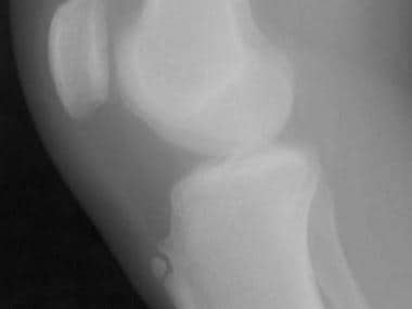 Radiograph of a patient who is skeletally mature. Note that the tibial tubercle is enlarged and that an ossicle is present, with an overlying bursa. Image courtesy of J Andy Sullivan, MD.
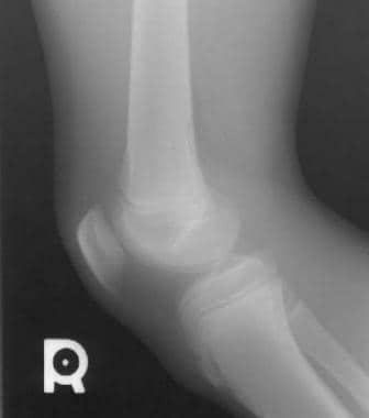 Radiograph of a patient who is skeletally immature. The tubercle is elongated and fragmented, and overlying soft-tissue swelling is present. Image courtesy of J Andy Sullivan, MD.
Soft-tissue edema in the region of the tibial tuberosity, with thickening and indistinct margins of the patellar tendon, enables the diagnosis of active Osgood-Schlatter disease with a high degree of confidence; usually, radiologic confirmation of this diagnosis is not necessary.
Accessory ossification centers may mimic findings in the late changes of Osgood-Schlatter disease. The radiographic differential diagnosis of multiple ossific opacities in the area of the anterior tibial tuberosity includes accessory ossification centers, which are normal variants, and late changes from a previous Osgood-Schlatter lesion.
-
Radiograph of a patient who is skeletally mature. Note that the tibial tubercle is enlarged and that an ossicle is present, with an overlying bursa. Image courtesy of J Andy Sullivan, MD.
-
Radiograph of a patient who is skeletally immature. The tubercle is elongated and fragmented, and overlying soft-tissue swelling is present. Image courtesy of J Andy Sullivan, MD.
